. 2010 Jul 8;5(7):e11489.
doi: 10.1371/journal.pone.0011489.

The human gonadotropin releasing hormone type I receptor is a functional intracellular GPCR expressed on the nuclear membrane

Affiliations

The human gonadotropin releasing hormone type I receptor is a functional intracellular GPCR expressed on the nuclear membrane

Michelle Re et al. PLoS One. .

Abstract

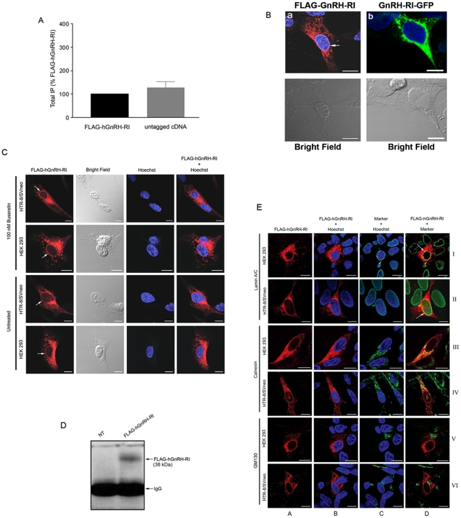
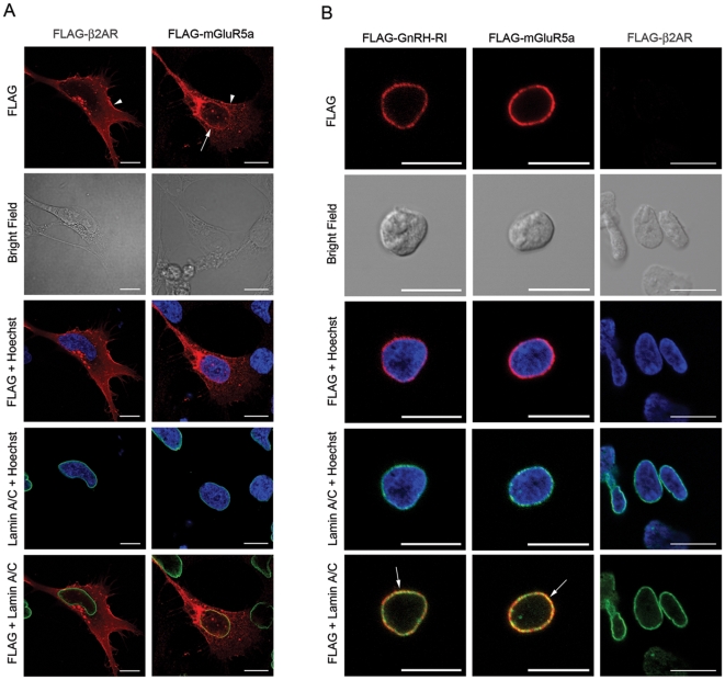
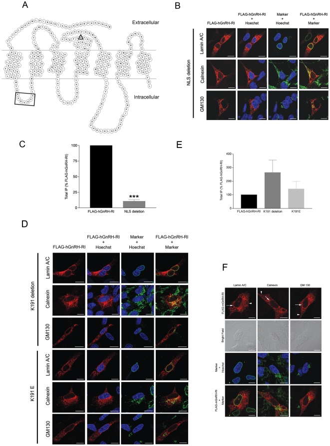
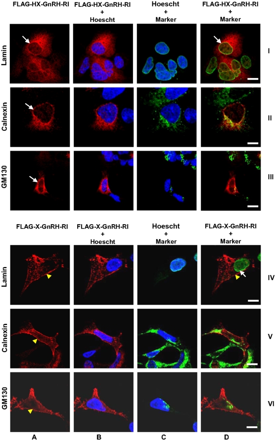
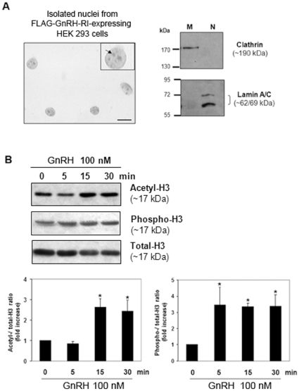
The mammalian type I gonadotropin releasing hormone receptor (GnRH-R) is a structurally unique G protein-coupled receptor (GPCR) that lacks cytoplasmic tail sequences and displays inefficient plasma membrane expression (PME). Compared to its murine counterparts, the primate type I receptor is inefficiently folded and retained in the endoplasmic reticulum (ER) leading to a further reduction in PME. The decrease in PME and concomitant increase in intracellular localization of the mammalian GnRH-RI led us to characterize the spatial distribution of the human and mouse GnRH receptors in two human cell lines, HEK 293 and HTR-8/SVneo. In both human cell lines we found the receptors were expressed in the cytoplasm and were associated with the ER and nuclear membrane. A molecular analysis of the receptor protein sequence led us to identify a putative monopartite nuclear localization sequence (NLS) in the first intracellular loop of GnRH-RI. Surprisingly, however, neither the deletion of the NLS nor the addition of the Xenopus GnRH-R cytoplasmic tail sequences to the human receptor altered its spatial distribution. Finally, we demonstrate that GnRH treatment of nuclei isolated from HEK 293 cells expressing exogenous GnRH-RI triggers a significant increase in the acetylation and phosphorylation of histone H3, thereby revealing that the nuclear-localized receptor is functional. Based on our findings, we conclude that the mammalian GnRH-RI is an intracellular GPCR that is expressed on the nuclear membrane. This major and novel discovery causes us to reassess the signaling potential of this physiologically and clinically important receptor.


Conflict of interest statement

Competing Interests: The authors have declared that no competing interests exist.

Figures

Figure 1
Figure 1. Effect of epitope tag on hGnRH-RI signaling and spatial localization of FLAG-hGnRH-RI.
(A) Inositol phosphate (IP) production in response to 100 nM Buserelin was assessed as described in “Materials and Methods”. Data represent three to six independent experiments performed in triplicate and normalized to FLAG-hGnRHRI ± S.E. (B, left) HTR-8/SVneo cells transfected with either FLAG-hGnRH-RI (a) subjected to indirect immunofluorescent staining using affinity purified rabbit anti-FLAG antibody followed by Alexa Fluor 568-conjugated anti-rabbit IgG (red) and counterstained with the nuclear dye, Hoechst (blue). Note the perinuclear localization of the FLAG-tagged hGnRH-RI (a, arrow); (B, right) HTR-8/SVneo cell transfected with GnRH-RI-GFP and counterstained with the nuclear dye, Hoechst (blue). Note the perinuclear localization of the GnRH-RI-GFP. (C) HTR-8/SVneo and HEK 293 cells transfected with FLAG-hGnRH-RI were subjected to indirect immunofluorescent staining using affinity purified rabbit anti-FLAG antibody followed by Alexa Fluor 568-conjugated anti-rabbit IgG (red) and counterstained with Hoechst (blue). Note the perinuclear localization of the FLAG-tagged hGnRH-RI seen in both cell lines (arrows). Bright field images are shown in the second column from the left. (D) Western blot analysis was performed on the lysates of nuclei isolated from HEK 293 cells overexpressing FLAG-GnRH-RI. The results reveal that the full length hGnRH-RI is expressed on the nuclei of HEK 293 cells. (E) Cells expressing FLAG-hGnRH-RI were subjected to indirect immunofluorescent staining for the receptor (red) as well as the nuclear membrane, endoplasmic reticulum and Golgi (all shown in green). Colocalization is seen as yellow staining. Column A: receptor alone; Column B: receptor + Hoescht; Column C: organelle marker (lamin A/C, calnexin or GM130) + Hoescht; Column D: receptor + organelle marker. Column letters and roman numerals used as a coordinate system. hGnRH-RI immunoreactivity co-localized with the nuclear marker, lamin A/C, as seen by the yellow staining (I-D, II-D, arrows) as well as with the endoplasmic reticulum marker, calnexin (III-D, IV-D). Less colocalization was seen between the receptor and the Golgi, as shown by less yellow staining (V-D, VI-D). Scale bar = 10 µm.
Figure 2
Figure 2. Spatial localization of FLAG-mGluR5a, FLAG-β2AR and FLAG-hGnRH-RI.
(A) HTR-8/SVneo cells transfected with either FLAG-mGluR5a or FLAG-β2AR were subjected to indirect immunofluorescent staining for the receptor (red) as well as the nuclear membrane marker lamin A/C (green). Cells were counterstained with Hoechst (blue). Note the perinuclear localization of the FLAG-tagged mGluR5a (arrow) and the plasma membrane localization of both FLAG-mGluR5a and FLAG-β2AR (arrowheads). Bright field images are shown in the second row from top. (B) Isolated nuclei from HEK293 cells expressing FLAG-hGnRH-RI, FLAG-mGluR5a or FLAG-β2AR were subjected to indirect immunofluorescent staining for the receptor (red) as well as the nuclear membrane marker lamin A/C (green). Cells were counterstained with Hoechst (blue). Note the colocalization of both FLAG-hGnRH-RI and FLAG-mGluR5a with the inner nuclear membrane marker lamin A/C (yellow, arrows). FLAG-β2AR was not localized to the nuclear membrane. Bright field images are shown in the second row from top. Scale bar = 10 µm.
Figure 3
Figure 3. Effect of putative NLS and lysine 191 hGnRH-RI mutants on receptor localization and signaling and spatial localization of FLAG-mGnRH-RI in HTR-8/SVneo cells.
(A) Schematic of the human GnRH-RI showing the positions of the lysine 191 residue (triangle) and the location of the putative NLS (square). (B) HTR-8/SVneo cells expressing FLAG-hGnRH-RI in which the putative NLS was deleted were subjected to indirect immunofluorescent staining for the receptor (red) as well as the markers for the nuclear membrane, endoplasmic reticulum and Golgi (shown in green). Cells were counterstained with Hoechst (blue). The NLS deletion mutant showed the same phenotype as the wild-type receptor, showing strong colocalization with lamin A/C and calnexin and less colocalization with GM130. (C) IP formation data represent seven independent experiments performed in triplicate and normalized to FLAG-hGnRHRI ± S.E. ***, p<0.001 versus IP formation of FLAG-hGnRH-RI. (D) HTR-8/SVneo cells expressing FLAG-hGnRH-RI in which lysine 191 was deleted or mutated to a glutamic acid residue (K191E) were subjected to indirect immunofluorescent staining for the receptor (red) as well as the markers for the nuclear membrane, endoplasmic reticulum and Golgi (shown in green). Cells were counterstained with Hoechst (blue). K191 deletion and K191E mutants showed the same phenotype as the wild-type receptor, showing strong colocalization with lamin A/C and calnexin and less colocalization with GM130. (E) IP formation data represent 6–7 independent experiments performed in triplicate and normalized to FLAG-hGnRHRI ± S.E. (F) HTR-8/SVneo cells expressing mGnRH-RI were subjected to indirect immunofluorescent staining for the receptor (red) as well as the markers for the nuclear membrane, endoplasmic reticulum and Golgi (shown in green). Cells were counterstained with Hoechst (blue). FLAG-mGnRH-RI showed similar perinuclear localization (arrows) to FLAG-hGnRH-RI with strong colocalization with lamin A/C and less colocalization with GM130. However, less colocalization was seen between the FLAG-mGnRH-RI and calnexin than was seen with the FLAG-hGnRH-RI. As well, there was evidence of plasma membrane localization (arrowheads) with FLAG-mGnRH-RI that was not seen with FLAG-hGnRH-RI. Bright field images are shown in the second row from top. Scale bar = 10 µm.
Figure 4
Figure 4. Spatial localization of FLAG-human-Xenopus (HX)-GnRH-RI and FLAG-Xenopus (X)-GnRH-RI in HEK 293 cells using organellar markers.
Cells expressing FLAG-GnRH-RI (HX and X) were subjected to indirect immunofluorescent staining for the receptor (red) as well as the nuclear membrane, endoplasmic reticulum and Golgi (all shown in green). Colocalization is seen as yellow staining. Column A: receptor alone; Column B: receptor + Hoescht; Column C: organelle marker (lamin A/C, calnexin or GM130) + receptor; Column D: receptor + organelle marker. Column letters and roman numerals used as a coordinate system. HX-GnRH-RI immunoreactivity co-localized with the nuclear marker, lamin A/C, as seen by the yellow staining (I-D, arrow) as well as the endoplasmic reticulum marker, calnexin (II-D). Less colocalization was seen between the receptor and the Golgi, as shown by less yellow staining (III-D). X-GnRH-RI immunoreactivity strongly localized to the plasma membrane (yellow arrowheads) and weakly in cytoplasm. No visual co-localization detected with lamin A/C (IV-D, arrow), calnexin (V-D) or Golgi (VI-D). Scale bar = 10 µm.
Figure 5
Figure 5. Effect of GnRH stimulation on Histone 3 acetylation/phosphorylation levels in isolated nuclei from HEK 293 transiently transfected with FLAG-GnRH-RI.
(A) Purity assessment of nuclei preparations used in functional assays by bright field microscopy and Western blotting. Left panel: Photomicrograph of a typical nuclei preparation following trypan blue staining. Scale bar = 10 µm. Insert: digital magnification of a single nucleus showing the presence of nucleoles (arrow). Right panel: Immunodetection of the membrane and nuclear markers clathrin and lamin A/C respectively in membrane and isolated nuclei fractions from FLAG-GnRH-R-transfected HEK 293 cells. (B) Time course of Histone H3-Lys9/Lys14 acetylation and -Ser10 phosphorylation in GnRH-stimulated nuclei by Western blot analysis. A representative Western blot is shown. Densitometric values of acetylated and phosphorylated Histone H3 levels were normalized to those of total Histone H3 and the relative intensity ratios were expressed in-fold increase over time zero. Data are means ± S.E.M of four independent experiments. *P<0.05 vs control time zero. Statistical analyses performed using one-way ANOVA followed by Dunnet's ad hoc test.

Similar articles

Cited by

References

    1. Cheng KW, Nathwani PS, Leung PC. Regulation of human gonadotropin-releasing hormone receptor gene expression in placental cells. Endocrinology. 2000;141:2340–2349. - PubMed
    1. Cavanagh PC, Dunk C, Pampillo M, Szereszewski JM, Taylor JE, et al. Gonadotropin-releasing hormone-regulated chemokine expression in human placentation. Am J Physiol Cell Physiol. 2009;297:C17–27. - PubMed
    1. Knollman PE, Conn PM. Multiple G proteins compete for binding with the human gonadotropin releasing hormone receptor. Arch Biochem Biophys. 2008;477:92–97. - PMC - PubMed
    1. Grosse R, Schmid A, Schoneberg T, Herrlich A, Muhn P, et al. Gonadotropin-releasing hormone receptor initiates multiple signaling pathways by exclusively coupling to G(q/11) proteins. J Biol Chem. 2000;275:9193–9200. - PubMed
    1. Maudsley S, Davidson L, Pawson AJ, Chan R, Lopez de Maturana R, et al. Gonadotropin-releasing hormone (GnRH) antagonists promote proapoptotic signaling in peripheral reproductive tumor cells by activating a Galphai-coupling state of the type I GnRH receptor. Cancer Res. 2004;64:7533–7544. - PubMed

Publication types