. 2010 Jul 7;5(7):e11468.
doi: 10.1371/journal.pone.0011468.

Mitochondrial DNA mutations induce mitochondrial dysfunction, apoptosis and sarcopenia in skeletal muscle of mitochondrial DNA mutator mice

Affiliations

Mitochondrial DNA mutations induce mitochondrial dysfunction, apoptosis and sarcopenia in skeletal muscle of mitochondrial DNA mutator mice

Asimina Hiona et al. PLoS One. .

Abstract

Background: Aging results in a progressive loss of skeletal muscle, a condition known as sarcopenia. Mitochondrial DNA (mtDNA) mutations accumulate with aging in skeletal muscle and correlate with muscle loss, although no causal relationship has been established.

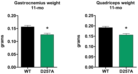
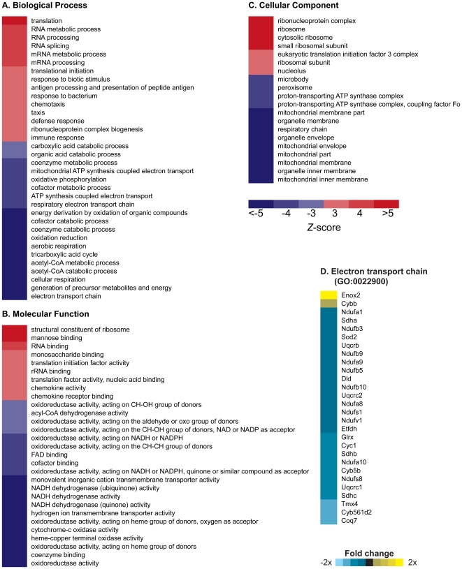
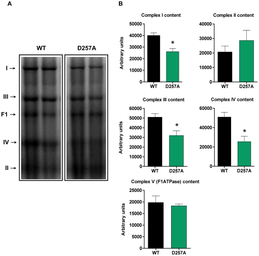
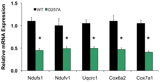
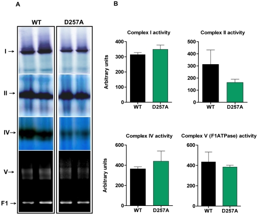
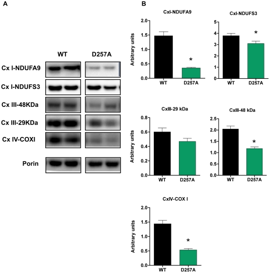
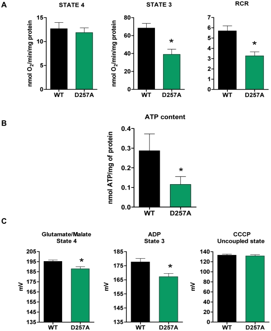
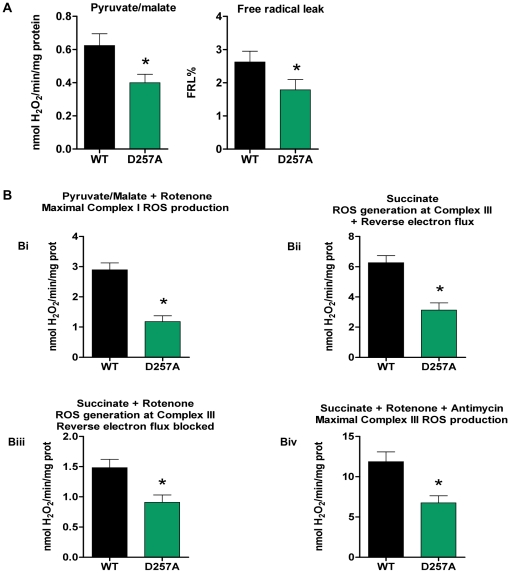
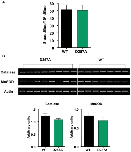
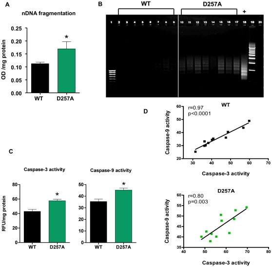
Methodology/principal findings: We investigated the relationship between mtDNA mutations and sarcopenia at the gene expression and biochemical levels using a mouse model that expresses a proofreading-deficient version (D257A) of the mitochondrial DNA Polymerase gamma, resulting in increased spontaneous mtDNA mutation rates. Gene expression profiling of D257A mice followed by Parametric Analysis of Gene Set Enrichment (PAGE) indicates that the D257A mutation is associated with a profound downregulation of gene sets associated with mitochondrial function. At the biochemical level, sarcopenia in D257A mice is associated with a marked reduction (35-50%) in the content of electron transport chain (ETC) complexes I, III and IV, all of which are partly encoded by mtDNA. D257A mice display impaired mitochondrial bioenergetics associated with compromised state-3 respiration, lower ATP content and a resulting decrease in mitochondrial membrane potential (Deltapsim). Surprisingly, mitochondrial dysfunction was not accompanied by an increase in mitochondrial reactive oxygen species (ROS) production or oxidative damage.

Conclusions/significance: These findings demonstrate that mutations in mtDNA can be causal in sarcopenia by affecting the assembly of functional ETC complexes, the lack of which provokes a decrease in oxidative phosphorylation, without an increase in oxidative stress, and ultimately, skeletal muscle apoptosis and sarcopenia.


Conflict of interest statement

Competing Interests: GCK and TAP were awarded United States patent 7,126,040 for the PolgD257A mouse model. TAP is a co-founder and scientific consultant for LifeGen Technologies, specializing in the application of DNA microarray analysis to analyzing nutraceutical interventions in aging. Dr. Barger of LifeGen Technologies was involved with analysis of gene expression data from microarray experiments. This does not alter the authors' adherence to all of the PLoS ONE policies on sharing data and materials.

Figures

Figure 1
Figure 1. D257A mice display significant skeletal muscle loss.
At 11 mo of age, gastrocnemius (n = 22 per group, *p<0.001) and quadriceps (n = 22 per group, *p<0.001) mean muscle mass in D257A mice is significantly decreased compared to age-matched WT. Error bars represent SEM.
Figure 2
Figure 2. Parametric Analysis of Gene Set Enrichment identifies Gene Ontology (GO) terms changed in gastrocnemius muscle of 13-month old D257A mice as compared to 13-month old WT mice.
(A, B, C) Terms shown are those significantly changed (p<0.01, FDR<0.01). Red indicates a GO term that was induced by mtDNA mutations (Z-score>0); blue indicates GO terms that were suppressed by mtDNA mutations (Z-score<0). n = 5 mice per group. (D) Genes listed within the GO term “electron transport chain” (Z-score = −8.89; p<0.0001) whose expression level significantly (p<0.01) increased (yellow) or decreased (blue) in D257A mice.
Figure 3
Figure 3. D257A mice display decreased content of ETC Complexes I, III and IV compared to WT.
(A) The total content of ETC complexes I, II, III, IV and the F1 domain of the ATPase from skeletal muscle of 11-mo old WT and D257A mice (n = 7 per group) was determined using Blue Native PAGE electrophoresis followed by staining with commassie blue stain. Proteins were separated according to molecular weight. Representative blots are depicted. (B) Statistical analysis of ETC complexes content. Arbitrary units represent densitometry values normalized to total protein loaded measured by the Bradford assay. Mean values ±SEM are shown. *p<0.01.
Figure 4
Figure 4. D257A mice display decreased mRNA expression of nuclear genes encoding components of ETC Complexes I, III and IV.
Relative mRNA expression of genes encoding components of the ETC complexes I, III and IV, including Ndufs1, Ndufv1, Uqcrc1, Cox6a2, and Cox7a1 was measured in the skeletal muscle from WT and D257A mice at 13 months of age by quantative RT-PCR (n = 5 per group). Mean values ±SEM are shown. *p<0.05.
Figure 5
Figure 5. No differences between WT and D257A mice in ETC complexes I, II, IV and F1-ATPase specific activity.
(A) The activity of ETC complexes I, II, IV and the F1 domain of the ATPase, in skeletal muscle of 11-mo old WT and D257A mice (n = 7 per group), was determined using Blue Native PAGE, followed by enzymatic colorimetric reactions performed on the gels. Representative blots are depicted. (B) Statistical analysis of ETC complexes specific activity. Arbitrary units represent activity densitometry values normalized to respective content densitometry values for each sample. No statistically significant differences were observed. Mean values ±SEM are shown.
Figure 6
Figure 6. D257A mice show decreased protein expression of both nuclear-encoded and mitochondrial-encoded ETC subunits.
(A) The content of selected nuclear-encoded (NDUFA9 and NDUFS3 subunits of Cx I, 29 kDa and 48 kDa subunits of Cx III), and mitochondrial-encoded subunits (COX I subunit of Cx IV) in skeletal muscle extracts of 11-mo old WT and D257A mice (n = 11 per group) were evaluated by Western Blotting. Representative blots are depicted. (B) Statistical analysis of ETC subunits protein expression. Arbitrary units represent densitometry values normalized to porin. Mean values ±SEM are shown. *p<0.05. Cx: complex.
Figure 7
Figure 7. Mitochondrial bioenergetics is compromised in D257A skeletal muscle leading to a drop in mitochondrial Δψm.
(A) We determined the effects of mtDNA mutations on O2 consumption of skeletal muscle mitochondria obtained from 11-mo old WT and D257A mice (n = 11 per group). Oxygen consumption was measured during state 4 (non-phosphorylative state) with pyruvate/malate as substrate, and during state 3 (phosphorylative state) with the addition of ADP. The respiratory control ratio (RCR), an index of mitochondrial coupling and metabolic activity, was calculated by dividing state 3 by state 4 respiration values. Mean values ±SEM are shown. *p<0.001. (B) We evaluated ATP content in skeletal muscle mitochondria of 11-mo old WT (n = 11) and D257A (n = 8) mice using a luciferin-luciferase based bioluminescence assay. Mean values ±SEM are shown. *p<0.05. (C) Changes in Δψm were followed qualitatively by monitoring the fluorescence of TMRM that accumulates in energized mitochondria of 13-mo old WT and D257A mice (n = 6 per group). Δψm was measured during both state 4 (non-phosphorylative state) with glutamate/malate as substrate and during state 3 (phosphorylative state) with the addition of ADP. Measurement of Δψm after addition of CCCP served as a control for TMRM loading in mitochondria. Mean values ±SEM are shown. *p<0.02.
Figure 8
Figure 8. D257A mitochondria produce fewer ROS in both main ROS generators (Complex I and Complex III).
(A) We measured H2O2 production using a sensitive fluorometric assay. Skeletal muscle mitochondria were obtained from 11-mo old, WT and D257A mice (n = 11 per group) and supplemented with pyruvate/malate as substrate for oxidative phosphorylation. Pyruvate/malate was used to study complex I ROS production (under near physiological conditions) which also represents total basal mitochondrial ROS production. Free radical leak percent (FRL%), an index of mitochondrial efficiency, was calculated by dividing the H2O2 value by twice the state 4 respiration value and the result was multiplied by 100 to give a % final value. Mean values ±SEM are shown. *p<0.05 (B) We used inhibitors of the ETC in order to study maximum rates of H2O2 production from complexes I and III, since they represent the main sites of ROS generation within the mitochondria (n = 11 per group). For complex I maximum rate (panel Bi) we used rotenone added to pyruvate/malate-supplemented mitochondria. For complex III maximum rate (panel Biv) we used antimycin A plus rotenone, added to succinate- supplemented mitochondria. We also used mitochondria supplemented with succinate alone in order to study complex III ROS production under near physiological conditions (panel Bii). In addition, some of the assays with succinate as substrate were performed in the presence of rotenone (panel Biii), in order to avoid the backwards flow of electrons to Complex I. Mean values ±SEM are shown. *p≤0.005.
Figure 9
Figure 9. Mitochondrial DNA oxidation levels and antioxidant enzyme mRNA expression remain unaltered in D257A skeletal muscle.
(A) We examined a marker of ROS-induced oxidative damage to DNA by assessing the levels of 8-oxodGuo in skeletal muscle mtDNA of 11-mo old WT and D257A mice (n = 11 per group), using HPLC with electrochemical detection. No statistically significant differences were detected. Mean values ±SEM are shown. (B) We measured Catalase and MnSOD mRNA expression in skeletal muscle extracts from 11-mo old WT (n = 7) and D257A (n = 8) mice by RT-PCR. Arbitrary units represent specific mRNA densitometry values normalized to actin mRNA densitometry values. No statistically significant differences were detected. Mean values ±SEM are shown.
Figure 10
Figure 10. Apoptosis is evident in D257A muscle by increased cytosolic DNA fragments, DNA laddering, and caspase activation.
(A) Cytosolic fractions from 11-mo old WT and D257A skeletal muscle (n = 11 per group) were prepared. Nuclear DNA fragmentation was quantified as the amount of mono- and oligo-nucleosomes present in the cytosol, using a sandwich ELISA. Mean values ±SEM are shown. *p<0.05. OD: optical density. (B) DNA from 13-mo old WT and D257A (n = 8 per group) mice was extracted and subjected to a DNA laddering-specific ligation PCR. PCR products were electrophoresed through 1% agarose gels and visualized under UV light for apoptosis-specific DNA ladders of ∼180–200 bp multiples. Lane 1: 100 bp molecular marker. Lanes 2–9: WT PCR products. Lanes 10–17: D257A PCR products. Lane 18: Positive control. Lane 19: 500 bp molecular marker. (C) Cytosolic fractions from 11-mo old WT and D257A skeletal muscle (n = 11 per group) were prepared. Caspase -3 and -9 activities were measured using a fluorometric protease assay kit which is based on detection of cleavage of the substrates DEVD-AFC or LEHD-AFC by caspase-3 and -9 respectively. Mean values ±SEM are shown. *p<0.002. RFU: raw fluorescent units. (D) Caspase-3 activity was correlated with caspase-9 activity in WT and D257A mice (n = 11 per group). Pearson correlation r values are shown in the top right corner. Correlations were significant for both genotypes; WT: r = 0.97, p<0.0001 and D257A: r = 0.8, p<0.003.

References

    1. Marcell TJ. Sarcopenia: causes, consequences, and preventions. J Gerontol A Biol Sci Med Sci. 2003;58:M911–916. - PubMed
    1. Roubenoff R. Sarcopenia and its implications for the elderly. Eur J Clin Nutr. 2000;54(Suppl 3):S40–47. - PubMed
    1. Janssen I, Baumgartner RN, Ross R, Rosenberg IH, Roubenoff R. Skeletal muscle cutpoints associated with elevated physical disability risk in older men and women. Am J Epidemiol. 2004;159:413–421. - PubMed
    1. Alexeyev MF, Ledoux SP, Wilson GL. Mitochondrial DNA and aging. Clin Sci (Lond) 2004;107:355–364. - PubMed
    1. Hiona A, Leeuwenburgh C. The role of mitochondrial DNA mutations in aging and sarcopenia: implications for the mitochondrial vicious cycle theory of aging. Exp Gerontol. 2008;43:24–33. - PMC - PubMed

Publication types

MeSH terms