. 2011 Sep;15(9):1999-2006.
doi: 10.1111/j.1582-4934.2010.01122.x. Epub 2010 Jul 12.

Radiosensitization of glioma cells by modulation of Met signalling with the hepatocyte growth factor neutralizing antibody, AMG102

Affiliations

Radiosensitization of glioma cells by modulation of Met signalling with the hepatocyte growth factor neutralizing antibody, AMG102

Ian M Buchanan et al. J Cell Mol Med. 2011 Sep.

Abstract

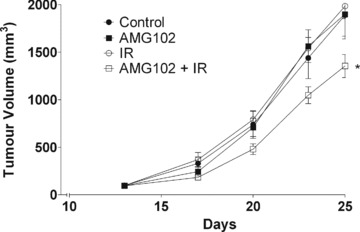
The hepatocyte growth factor (HGF)/Met signalling pathway is up-regulated in many cancers, with downstream mediators playing a role in DNA double strand break repair. Previous studies have shown increased radiosensitization of tumours through modulation of Met signalling by genetic methods. We investigated the effects of the anti-HGF monoclonal antibody, AMG102, on the response to ionizing radiation in a model of glioblastoma multiforme in vitro and in vivo. Radiosensitivity was evaluated in vitro in the U-87 MG human glioma cell line. Met activation was measured by Western blot, and the effect on survival following radiation was evaluated by clonogenic assay. Mechanism of cell death was evaluated by apoptosis and mitotic catastrophe assays. DNA damage was quantitated by γH2AX foci and neutral comet assay. Growth kinetics of subcutaneous tumours was used to assess the effects of AMG102 on in vivo tumour radiosensitivity. AMG102 inhibited Met activation after irradiation. An enhancement of radiation cell killing was shown with no toxicity using drug alone. Retention of γH2AX foci at 6 and 24 hrs following the drug/radiation combination indicated an inhibition of DNA repair following radiation, and comet assay confirmed DNA damage persisting over the same duration. At 48 and 72 hrs following radiation, a significant increase of cells undergoing mitotic catastrophe was seen in the drug/radiation treated cells. Growth of subcutaneous tumours was slowed in combination treated mice, with an effect that was greater than additive for each modality individually. Modulation of Met signalling with AMG102 may prove a novel radiation sensitizing strategy. Our data indicate that DNA repair processes downstream of Met are impaired leading to increased cell death through mitotic catastrophe.


Figures

Fig 1
Fig 1
The influence of AMG102 on Met activation in U-87 MG cells. Cells were exposed to AMG102 (1 μg/ml) for 24 hrs, irradiated (2 Gy) and collected at 1, 6 and 24 hr time-points.
Fig 2
Fig 2
The influence of AMG102 on U-87 MG tumour cell radiosensitivity. U-87 MG cells were exposed to AMG102 or IgG2 control (1 μg/ml) for 16 hrs and exposed to between 0 and 8 Gy IR. Colony-forming efficiency was determined 10–14 days later and survival curves generated after normalizing for cytotoxicity due to AMG102 alone. PE: plating efficiency; DEF0.1: dose enhancement factor at 10% surviving fraction; •: IgG2 control; ▪: AMG102. n = 3; points: mean; bars: ±S.E.
Fig 3
Fig 3
The influence of AMG102 on mechanism of cell death in U-87 MG cells. (A) Apoptosis. U-87 MG cells were exposed to AMG102 (1 μg/ml), irradiated (2 Gy) and collected at 24 and 72 hr time-points for analysis by flow cytometry. Apoptotic Index,% annexin-V + 7-AAD+ cells. n = 3; columns: mean; bars: ±S.E. (B) Mitotic catastrophe. U-87 MG cells were exposed to AMG102 (1 μg/ml) for 24 hrs, irradiated (2 Gy) and fixed at 24, 48 and 72 hr time-points. Immunofluorescence staining was performed. Nuclei from each replicate scored three separate times in the same 150–200 cells, with the average taken. Mitotic Catastrophe Index,% cells present with ≥2 nuclear fragments. n = 3; columns: mean; bars: ±S.E.; * P = 0.0310, ** P = 0.0026 (unpaired t-test, IR versus AMG102 + IR).
Fig 3
Fig 3
The influence of AMG102 on mechanism of cell death in U-87 MG cells. (A) Apoptosis. U-87 MG cells were exposed to AMG102 (1 μg/ml), irradiated (2 Gy) and collected at 24 and 72 hr time-points for analysis by flow cytometry. Apoptotic Index,% annexin-V + 7-AAD+ cells. n = 3; columns: mean; bars: ±S.E. (B) Mitotic catastrophe. U-87 MG cells were exposed to AMG102 (1 μg/ml) for 24 hrs, irradiated (2 Gy) and fixed at 24, 48 and 72 hr time-points. Immunofluorescence staining was performed. Nuclei from each replicate scored three separate times in the same 150–200 cells, with the average taken. Mitotic Catastrophe Index,% cells present with ≥2 nuclear fragments. n = 3; columns: mean; bars: ±S.E.; * P = 0.0310, ** P = 0.0026 (unpaired t-test, IR versus AMG102 + IR).
Fig 4
Fig 4
The influence of AMG102 on radiation-induced DNA damage in U-87 MG cells. (A) γH2AX foci. U-87 MG cells were exposed to AMG102 or vehicle control IgG2 (1 μg/ml) for 24 hrs, irradiated (2 Gy), and fixed at 1, 6 and 24 hr time-points. Immunofluorescence staining was performed. Nuclear foci from each replicate counted three separate times in the same 50 cells, with the average taken. n = 3; columns: mean; bars: ±S.E.; *P = 0.0002, **P < 0.0001 (unpaired t-test, IR versus AMG102 + IR). (B) Neutral comet assay. U-87 MG cells were exposed to AMG102 (1 μg/ml) for 24 hrs, irradiated (10 Gy) and collected at 0, 1, 6 and 24 hr time-points. 25–50 cells analysed per condition. Olive Tail Moment = (Tail mean – Head mean) × Tail%DNA/100; •: untreated control; ▪: AMG102; ○: IR; □: AMG102 + IR. Representative figure, repeated three times; points: mean; bars: ±S.E.; *,** P < 0.0001 (unpaired t-test, IR versus AMG102 + IR).
Fig 4
Fig 4
The influence of AMG102 on radiation-induced DNA damage in U-87 MG cells. (A) γH2AX foci. U-87 MG cells were exposed to AMG102 or vehicle control IgG2 (1 μg/ml) for 24 hrs, irradiated (2 Gy), and fixed at 1, 6 and 24 hr time-points. Immunofluorescence staining was performed. Nuclear foci from each replicate counted three separate times in the same 50 cells, with the average taken. n = 3; columns: mean; bars: ±S.E.; *P = 0.0002, **P < 0.0001 (unpaired t-test, IR versus AMG102 + IR). (B) Neutral comet assay. U-87 MG cells were exposed to AMG102 (1 μg/ml) for 24 hrs, irradiated (10 Gy) and collected at 0, 1, 6 and 24 hr time-points. 25–50 cells analysed per condition. Olive Tail Moment = (Tail mean – Head mean) × Tail%DNA/100; •: untreated control; ▪: AMG102; ○: IR; □: AMG102 + IR. Representative figure, repeated three times; points: mean; bars: ±S.E.; *,** P < 0.0001 (unpaired t-test, IR versus AMG102 + IR).
Fig 5
Fig 5
The influence of AMG102 on U-87 MG tumour growth. U-87 MG subcutaneous xenograft tumours grown to ≍100 mm3 before randomization to four groups: untreated controls, AMG102, IR, AMG102 + IR. AMG102 (15 μg) was injected i.p. and tumours irradiated (3 Gy) 16 hrs later with animals shielded in custom lead jigs. Tumour volume: [L × W2]/2; •: untreated control; ▪: AMG102; ○: IR; □: AMG102 + IR. Representative figure, repeated two times; points: mean; bars: ±S.E.; * P < 0.0241 (unpaired t-test, AMG102 + IR versus all other groups).

References

    1. Pazdur R, Wagman LD, Camphausen KA, et al. Oncology, editor. 11 ed. Lawrence, KS, USA: Medica; 2008. Cancer management: a multidiciplinary approach, medical, surgical, & radiation oncology.
    1. Cooper C, Park M, Blair D, et al. Molecular cloning of a new transforming gene from a chemically transformed human cell line. Nature. 1984;311:29–33. - PubMed
    1. Stoker M, Gherardi E, Perryman M, et al. Scatter factor is a fibroblast-derived modulator of epithelial cell mobility. Nature. 1987;327:239–42. - PubMed
    1. Nakamura T, Nishikawa T, Hagiya M, et al. Molecular cloning and expression of human hepatocyte growth factor. Nature. 1989;342:440–3. - PubMed
    1. Weidner KM, Arakaki N, Hartmann G, et al. Evidence for the identity of human scatter factor and human hepatocyte growth factor. Proc Natl Acad Sci USA. 1991;88:7001–5. - PMC - PubMed

Publication types

MeSH terms