. 2010 Aug;14(8):2085-93.
doi: 10.1111/j.1582-4934.2010.01129.x. Epub 2010 Jul 13.

Telocytes in human epicardium

Affiliations

Telocytes in human epicardium

L M Popescu et al. J Cell Mol Med. 2010 Aug.

Abstract

The existence of the epicardial telocytes was previously documented by immunohistochemistry (IHC) or immunofluorescence. We have also demonstrated recently that telocytes are present in mice epicardium, within the cardiac stem-cell niches, and, possibly, they are acting as nurse cells for the cardiomyocyte progenitors. The rationale of this study was to show that telocytes do exist in human (sub)epicardium, too. Human autopsy hearts from 10 adults and 15 foetuses were used for conventional IHC for c-kit/CD117, CD34, vimentin, S-100, τ, Neurokinin 1, as well as using laser confocal microscopy. Tissue samples obtained by surgical biopsies from 10 adults were studied by digital transmission electron microscopy (TEM). Double immunolabelling for c-kit/CD34 and, for c-kit/vimentin suggests that in human beings, epicardial telocytes share similar immunophenotype features with myocardial telocytes. The presence of the telocytes in human epicardium is shown by TEM. Epicardial telocytes, like any of the telocytes are defined by telopodes, their cell prolongations, which are very long (several tens of μm), very thin (0.1-0.2 μm, below the resolving power of light microscopy) and with moniliform configuration. The interconnected epicardial telocytes create a 3D cellular network, connected with the 3D network of myocardial telocytes. TEM documented that telocytes release shed microvesicles or exocytotic multivesicular bodies in the intercellular space. The human epicardial telocytes have similar phenotype (TEM and IHC) with telocytes located among human working cardiomyocyte. It remains to be established the role(s) of telocytes in cardiac renewing/repair/regeneration processes, and also the pathological aspects induced by their 'functional inhibition', or by their variation in number. We consider telocytes as a real candidate for future developments of autologous cell-based therapy in heart diseases.


Figures

Fig 1
Fig 1
(A and B) Confocal microscopy. c-kit+ cells in human subepicardial area. (A). Gallery of LSM (laser scanning microscopy) images obtained at 0.529 μM steps showing the spatial distribution of c-kit+ cells with long processes under mesothelial cells (arrows) in human myocardium. (B) Maximum projection of LSM images shown in (A). Mesothelial cells are indicated with arrows. (C) TEM: one telocyte is located near the mesothelial cells of the same human epicardium.
Fig 2
Fig 2
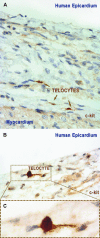
Immunostaining for c-kit in human epicardium. (A) c-kit positivity of telocytes is more evident at the level of the cell bodies in the deeper epicardial layer. Original magnification, 40×. (B) Immunostaining revealing a c-kit+ cell near epicardial surface. Note that immunoreactivity is not restricted to the cell body, and clearly reveals the emergence of telopodes. Original magnification, 60×. (C) Higher magnification of the telocytes presented in Fig. 2B. Telopodes are running out in opposite directions and their thickness at the advent is rather discrete. The staining is concomitant on cell body and cell prolongations. Original magnification, 100×.
Fig 3
Fig 3
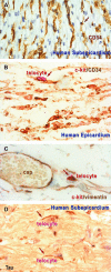
(A) Immunostaining for CD34 (brown). CD34 positivity might be expressed by telocytes and endothelial cells. (B) Localization of double positive cells for c-Kit (red) and CD34 (brown). (C) Human epicardium, deep layer. Vimentin positivity is stronger than c-Kit positivity by sandwich method. (D) Cells expressing τ protein (arrows). (A–D) Original magnification, 40×.
Fig 4
Fig 4
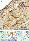
(A) reveals a cluster of three cells positive for S-100 (arrows) distributed in human subepicardium. (B) Immunostaining for CD57 (NK1) in human subepicardium. Thin cellular processes are stained in the vicinity of capillaries. Original magnification, 40×.
Fig 5
Fig 5
Digital coloured TEM images show telocytes (blue) in the human subepicardium, bordering the peripheral cardiomyocytes (CM, highlighted in brown). (A) TEM image of a 20 μm long telocyte with three telopodes, illustrating the distinctive dichotomous pattern of branching (arrows). (B) The thin, moniliform telopodes (length for the convoluted telopode –30 μm, upper telopode –38 μm) overlap each other in the periphery of myocardium creating a separation leaf. E-elastic fibres, coll-collagen fibres. Scale bar –5 μm.
Fig 6
Fig 6
Digitally coloured electron micrographs show telopodes (blue) and shed microvesicles (purple) in the extracellular matrix in subepicardium. (A) The telopode has a lacunar aspect and microvesicles seem to be released in the extracellular matrix or endocytosed into cytoplasmic pockets (arrows). (B) A telopode delivers few microvesicles (arrow) in the vicinity of a nerve fibre (green). Few caveolae (asterisks) are visible on plasma membrane of the telopode. (C) A multivesicular body (arrow) emerges from a telopode and discards its microvesicles (about 100 nm diameter) for another telopode. Arrowhead indicates a small attachment plaque connecting the telopode to extracellular matrix. Scale bar –0.5 μm.
Fig 7
Fig 7
Electron micrographs of human subepicardium show that telopodes organized in a labyrinthine system connect nerves (green coloured in A and C), monocytes (B) and adipocytes (light brown, C). Cardiomyocytes (CM) are coloured in brown. Scale bar –2 μm.

References

    1. Zhou B, Pu WT. More than a cover: epicardium as a novel source of cardiac progenitor cells. Regen Med. 2008;3:633–5. - PubMed
    1. Manner J. Does the subepicardial mesenchyme contribute myocardioblasts to the myocardium of the chick embryo heart? A quail-chick chimera study tracing the fate of the epicardial primordium. Anat Rec. 1999;255:212–26. - PubMed
    1. Lie-Venema H, Van Den Akker NM, Bax NA, et al. Origin, fate, and function of epicardium-derived cells (EPDCs) in normal and abnormal cardiac development. ScientificWorldJournal. 2007;7:1777–98. - PMC - PubMed
    1. Zhang F, Pasumarthi KB. Ultrastructural and immunocharacterization of undifferentiated myocardial cells in the developing mouse heart. J Cell Mol Med. 2007;11:552–60. - PMC - PubMed
    1. Zhou B, Ma Q, Rajagopal S, Wu SM, et al. Epicardial progenitors contribute to the cardiomyocyte lineage in the developing heart. Nature. 2008;454:109–13. - PMC - PubMed

MeSH terms