

Gonadotropin-releasing hormone type II antagonist induces apoptosis in MCF-7 and triple-negative MDA-MB-231 human breast cancer cells in vitro and in vivo
- PMID: 20630060
- PMCID: PMC2949636
- DOI: 10.1186/bcr2606
Gonadotropin-releasing hormone type II antagonist induces apoptosis in MCF-7 and triple-negative MDA-MB-231 human breast cancer cells in vitro and in vivo
Abstract
Introduction: Triple-negative breast cancer does not express estrogen and progesterone receptors, and no overexpression/amplification of the HER2-neu gene occurs. Therefore, this subtype of breast cancer lacks the benefits of specific therapies that target these receptors. Today chemotherapy is the only systematic therapy for patients with triple-negative breast cancer. About 50% to 64% of human breast cancers express receptors for gonadotropin-releasing hormone (GnRH), which might be used as a target. New targeted therapies are warranted. Recently, we showed that antagonists of gonadotropin-releasing hormone type II (GnRH-II) induce apoptosis in human endometrial and ovarian cancer cells in vitro and in vivo. This was mediated through activation of stress-induced mitogen-activated protein kinases (MAPKs) p38 and c-Jun N-terminal kinase (JNK), followed by activation of proapoptotic protein Bax, loss of mitochondrial membrane potential, and activation of caspase-3. In the present study, we analyzed whether GnRH-II antagonists induce apoptosis in MCF-7 and triple-negative MDA-MB-231 human breast cancer cells that express GnRH receptors. In addition, we ascertained whether knockdown of GnRH-I receptor expression affects GnRH-II antagonist-induced apoptosis and apoptotic signaling.
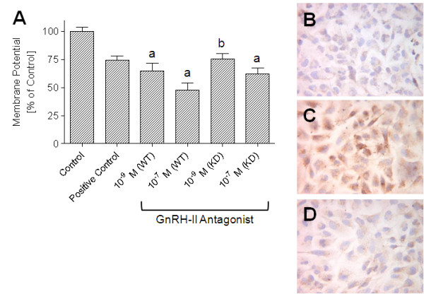
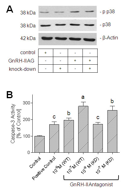
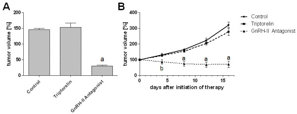
Methods: Induction of apoptosis was analyzed by measurement of the loss of mitochondrial membrane potential. Apoptotic signaling was measured with quantification of activated MAPK p38 and caspase-3 by using the Western blot technique. GnRH-I receptor protein expression was inhibited by using the antisense knockdown technique. In vivo experiments were performed by using nude mice bearing xenografted human breast tumors.
Results: We showed that treatment of MCF-7 and triple-negative MDA-MB-231 human breast cancer cells with a GnRH-II antagonist results in apoptotic cell death in vitro via activation of stress-activated MAPK p38 and loss of mitochondrial membrane potential. In addition, we showed GnRH-II antagonist-induced activation of caspase-3 in MDA-MB-231 human breast cancer cells. After knockdown of GnRH-I receptor expression, GnRH-II antagonist-induced apoptosis and apoptotic signaling was only slightly reduced, indicating that an additional pathway mediating the effects of GnRH-II antagonists may exist. The GnRH-I receptor seems not to be the only target of GnRH-II antagonists. The antitumor effects of the GnRH-II antagonist could be confirmed in nude mice. The GnRH-II antagonist inhibited the growth of xenotransplants of human breast cancers in nude mice completely, without any apparent side effects.
Conclusions: GnRH-II antagonists seem to be suitable drugs for an efficacious and less-toxic endocrine therapy for breast cancers, including triple-negative breast cancers.
Figures
Similar articles
-
Targeted chemotherapy for triple-negative breast cancers via LHRH receptor.Oncol Rep. 2011 May;25(5):1481-7. doi: 10.3892/or.2011.1188. Epub 2011 Feb 17. Oncol Rep. 2011. PMID: 21331448
-
Agonists and antagonists of GnRH-I and -II reduce metastasis formation by triple-negative human breast cancer cells in vivo.Breast Cancer Res Treat. 2011 Dec;130(3):783-90. doi: 10.1007/s10549-011-1358-9. Epub 2011 Jan 30. Breast Cancer Res Treat. 2011. PMID: 21279682
-
GnRH-II antagonists induce apoptosis in human endometrial, ovarian, and breast cancer cells via activation of stress-induced MAPKs p38 and JNK and proapoptotic protein Bax.Cancer Res. 2009 Aug 15;69(16):6473-81. doi: 10.1158/0008-5472.CAN-08-4657. Epub 2009 Jul 28. Cancer Res. 2009. PMID: 19638591
-
Role of gonadotropin-releasing hormone (GnRH) in ovarian cancer.Reprod Biol Endocrinol. 2003 Oct 7;1:65. doi: 10.1186/1477-7827-1-65. Reprod Biol Endocrinol. 2003. PMID: 14594454 Free PMC article. Review.
-
GnRH antagonists in the treatment of gynecological and breast cancers.Endocr Relat Cancer. 2003 Jun;10(2):291-9. doi: 10.1677/erc.0.0100291. Endocr Relat Cancer. 2003. PMID: 12790790 Review.
Cited by
-
Treatment of Breast Cancer With Gonadotropin-Releasing Hormone Analogs.Front Oncol. 2019 Oct 1;9:943. doi: 10.3389/fonc.2019.00943. eCollection 2019. Front Oncol. 2019. PMID: 31632902 Free PMC article. Review.
-
Role of Gonadotropin-Releasing Hormone (GnRH) in Ovarian Cancer.Cells. 2021 Feb 18;10(2):437. doi: 10.3390/cells10020437. Cells. 2021. PMID: 33670761 Free PMC article. Review.
-
Effect of GnRH-II on the ESC proliferation, apoptosis and VEGF secretion in patients with endometriosis in vitro.Int J Clin Exp Pathol. 2013 Oct 15;6(11):2487-96. eCollection 2013. Int J Clin Exp Pathol. 2013. PMID: 24228111 Free PMC article.
-
Dissecting the Hormonal Signaling Landscape in Castration-Resistant Prostate Cancer.Cells. 2021 May 7;10(5):1133. doi: 10.3390/cells10051133. Cells. 2021. PMID: 34067217 Free PMC article. Review.
-
Expression and Role of Gonadotropin-Releasing Hormone 2 and Its Receptor in Mammals.Front Endocrinol (Lausanne). 2017 Dec 11;8:269. doi: 10.3389/fendo.2017.00269. eCollection 2017. Front Endocrinol (Lausanne). 2017. PMID: 29312140 Free PMC article. Review.
References
-
- Ferlay J, Bray F, Pisani P, Parkin DM. GLOBOCAN 2000: cancer incidence, mortality and prevalence worldwide, version 1.0. IARC CancerBase No. 5. IARC Press, Lyon; 2001.
-
- Konecny G, Pauletti G, Pegram M, Untch M, Dandekar S, Aguilar Z, Wilson C, Rong HM, Bauerfeind I, Felber M, Wang HJ, Beryt M, Seshadri R, Hepp H, Slamon DJ. Quantitative association between HER-2/neu and steroid hormone receptors in hormone receptor-positive primary breast cancer. J Natl Cancer Inst. 2003;95:142–153. doi: 10.1093/jnci/95.2.142. - DOI - PubMed
-
- Nielsen TO, Hsu FD, Jensen K, Cheang M, Karaca G, Hu Z, Hernandez-Boussard T, Livasy C, Cowan D, Dressler L, Akslen LA, Ragaz J, Gown AM, Gilks CB, van de Rijn M, Perou CM. Immunohistochemical and clinical characterization of the basal-like subtype of invasive breast carcinoma. Clin Cancer Res. 2004;10:5367–5374. doi: 10.1158/1078-0432.CCR-04-0220. - DOI - PubMed
Publication types
MeSH terms
Substances
LinkOut - more resources
Full Text Sources
Other Literature Sources
Research Materials
Miscellaneous
