. 2010 Nov 1;53(2):450-9.
doi: 10.1016/j.neuroimage.2010.06.072. Epub 2010 Jul 11.

Cerebral cortical folding analysis with multivariate modeling and testing: Studies on gender differences and neonatal development

Affiliations

Cerebral cortical folding analysis with multivariate modeling and testing: Studies on gender differences and neonatal development

Suyash P Awate et al. Neuroimage. .

Abstract

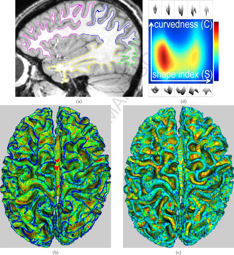
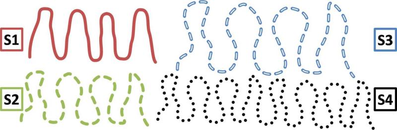
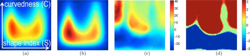
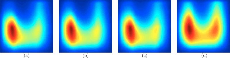
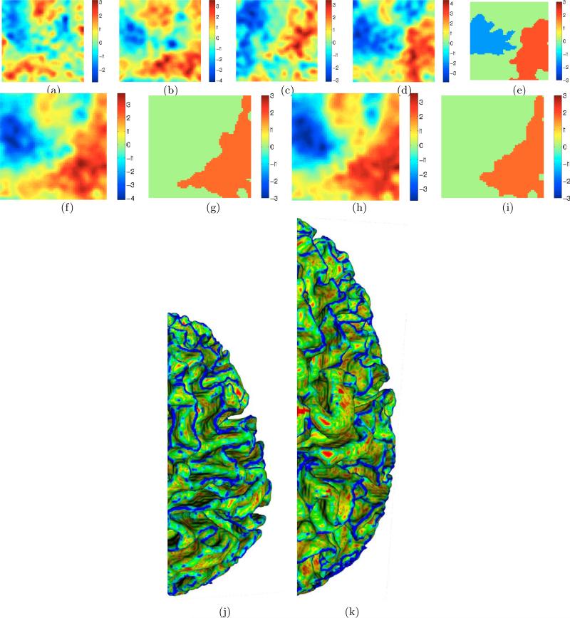
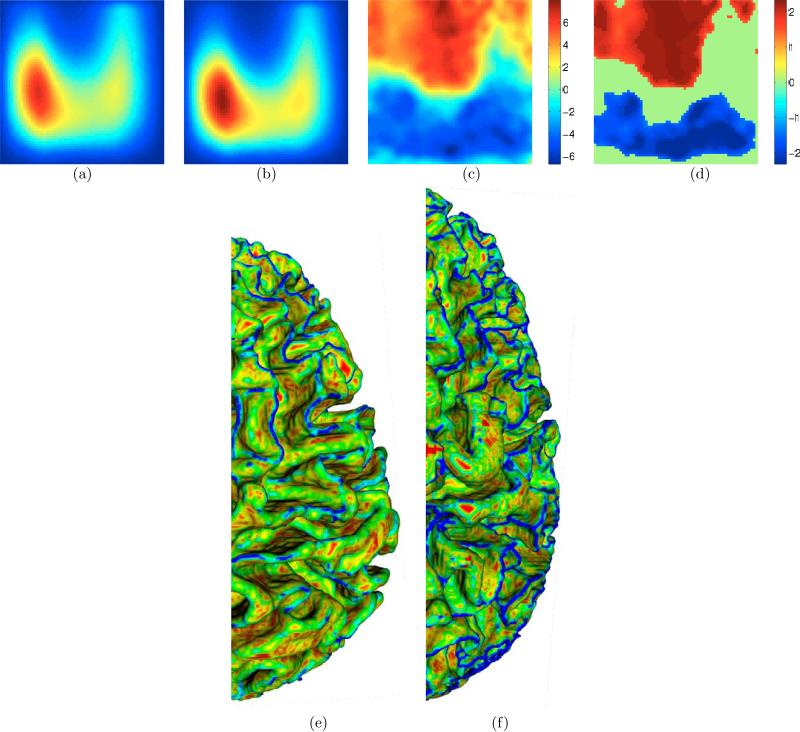
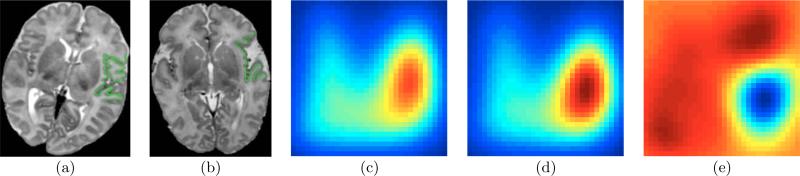
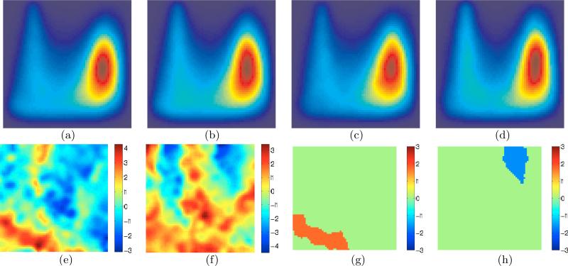
This paper presents a novel statistical framework for human cortical folding pattern analysis that relies on a rich multivariate descriptor of folding patterns in a region of interest (ROI). The ROI-based approach avoids problems faced by spatial normalization-based approaches stemming from the deficiency of homologous features between typical human cerebral cortices. Unlike typical ROI-based methods that summarize folding by a single number, the proposed descriptor unifies multiple characteristics of surface geometry in a high-dimensional space (hundreds/thousands of dimensions). In this way, the proposed framework couples the reliability of ROI-based analysis with the richness of the novel cortical folding pattern descriptor. This paper presents new mathematical insights into the relationship of cortical complexity with intra-cranial volume (ICV). It shows that conventional complexity descriptors implicitly handle ICV differences in different ways, thereby lending different meanings to "complexity". The paper proposes a new application of a nonparametric permutation-based approach for rigorous statistical hypothesis testing with multivariate cortical descriptors. The paper presents two cross-sectional studies applying the proposed framework to study folding differences between genders and in neonates with complex congenital heart disease. Both studies lead to novel interesting results.


Figures

Figure 1
Figure 1
(a) A sagittal slice of a head MR image overlapped with the cortical surface M. (b) Curvedness values C(m) painted on M (red→blue ≡ zero→high). In this figure, red areas are almost flat and blue areas are highly curved. (c) Shape-index values S(m) ∈ [−1, 1] painted on M (red→blue ≡ −1 → 1). In this figure, red/yellow areas are concave, blue/cyan areas are convex, and green areas are saddles. (d) Proposed descriptor PM(C,S) (blue→red ≡ 0 → 1 probability; colormap shown on right). For all plots of PM(C,S) in this paper: horizontal axis ≡ S, vertical axis ≡ C. The illustrations of local surface patches, at the top and bottom of this figure, depict patches for the entire range of possible S values; corresponding patches at the top and bottom have the same S value; all patches at the top have the same C value that is higher than the C value for all the patches at the bottom; the left half of the domain comprises concave patches, while the right half comprises convex patches.
Figure 2
Figure 2
What does “complexity” mean when volumes differ: issues of scale and replication. S1 and S2 occupy the same volume (i.e. volume of their convex hulls); S2 is more complex than S1. S3 and S4 occupy larger volumes than S2. S3 enlarges/scales the folds in S2. S4 replicates the folds in S2. How do we compare the complexities of (i) S3 and S2 and (ii) S4 and S2 ?
Figure 3
Figure 3
Validation : a simulated cross-sectional study between the group of cortical surfaces (interface between GM and WM) of 20 BrainWeb [Aubert-Broche et al. (2006)] images and a second group created by moderately smoothing the 20 BrainWeb surfaces. For all plots in this paper, horizontal axis ≡ S, vertical axis ≡ C, coordinates for the bottom left corner : (C, S) = (cmin, −1); bottom right corner : (C, S) = (cmin, 1); top left corner : (C, S) = (cmax, −1). (a)-(b) Mean of the multivariate surface descriptors PMn(C,S) for the n = 1, . . . , 20 original and smoothed surfaces, respectively, as proposed in Section 2.1; red≡high and blue≡low values. (c) The t-statistic map for the original and smoothed surfaces; t > 0 ⇒ Poriginal > Psmoothed. Expectedly, the map indicates that the original surfaces have larger mass (red/yellow) in high-curvedness regions as compared to the smoothed surfaces. (d) The significant locations (p < 0.05) produced via permutation testing [Fisher (1935); Nichols and Holmes (2002)]. For all plots in this paper, p values (corrected; permutation testing) for significant locations/clusters are visualized by coloring them by the associated z score [Papoulis and Pillai (2001)], e.g. z(p = 0.05) = 1.65, z(p = 0.005) = 2.58.
Figure 4
Figure 4
Validation : measuring the fraction of surface area of the cortical GM-WM surface comprising concave surface patches (predominantly sulci) in 50 healthy adults. (a)-(d) Proposed surface descriptors for the frontal, parietal, temporal, and occipital lobes, respectively. The asymmetry between the left and right halves, i.e. concave and convex patches, is consistent with published clinical studies [Van-Essen and Drury (1997); Zilles et al. (1988)]. Note the reduced left-right asymmetry in (d) relative to (a)-(c).
Figure 5
Figure 5
(a)-(d) Student's t statistics for a cross-sectional study with Preplication (positive t ≡ larger value in males) for frontal, parietal, temporal, and occipital lobes, respectively, in the left hemisphere. Similar patterns exist for lobes in right hemisphere. (e) Significant clusters (via permutation testing) for left occipital lobe. (f ),(h) t statistics and (g),(i) significant clusters (via permutation testing) for Preplication for left hemisphere (4 lobes) and whole brain (8 lobes), respectively. A similar pattern exists for the right hemisphere. Note the larger mass (red) for males in the low-curvedness regions (bottom half) and larger mass (red) for males in the convex regions (right half). (j)-(k) Selected smaller-than-average female and larger-than-average male brains, respectively, painted by C values (red→blue ≡ zero→high) to enable/accentuate visualization of the differences. Thus, based on the meaning of complexity ingrained in Preplication, the female cortex appears more “complex”, i.e. more blue/cyan. Compare this with Figure 6(e-f).
Figure 6
Figure 6
(a)-(b) Average of Pscale for males and females, respectively, for the left frontal lobe. (c)-(d) t statistics (positive t ≡ larger value in males) and significant locations (via permutation testing), respectively, for the left frontal lobe. Similar patterns exist for all other lobes. Note the larger mass (red) for males in the high-curvedness (top half) regions. (e)-(f) Selected smaller-than-average female and larger-than-average male brains, respectively, adjusted for ICV and painted by C values (red→blue ≡ zero→high) to enable/accentuate visualization of the differences. Thus, based on the meaning of complexity ingrained in Pscale, the female brain appears less “complex”, i.e. more red/yellow. Compare this with Figure 5(j-k).
Figure 7
Figure 7
(a)-(b) Example MR images of the normal (mature, “closed”, more folded) and farthest-from-normal (immature, “open”, less folded) operculums, respectively, overlapped with the extracted cortical surfaces. (c)-(d) Average cortical folding descriptors Paverage normal(C, S) and Paverage abnormal(C, S) for 2 closest-to-normal and 4 significantly-abnormal operculums, respectively, chosen by a medical expert based on a subjective clinical scoring protocol [Licht et al. (2009)]. (e) Paverage normal(C, S) − Paverage abnormal(C, S); blue ≡ negative and red ≡ positive values.
Figure 8
Figure 8
(a)-(b) Average of cortical folding descriptors P(C, S) in right and left operculums, respectively, for 29 neonates with HLHS. (c)-(d) Average of the descriptors in right and left operculums, respectively, for 13 neonates with TGA. (e)-(f) t-statistic maps; t > 0 ⇒ PHLHS > PTGA. for right and left operculums, respectively. (g)-(h) Significant clusters (via permutation testing) for right and left operculums, respectively.

References

    1. Armstrong E, Schleicher A, Omran H, Curtis M, Zilles K. The ontogeny of human gyrification. Cerebral Cortex. 1995;5:56–63. - PubMed
    1. Ashburner J, Friston K. Voxel-based morphometry - the methods. NeuroImage. 2000;11(6):805–821. - PubMed
    1. Aubert-Broche B, Collins D, Evans A. Twenty new digital brain phantoms for creation of validation image data bases. IEEE Trans. Med. Imag. 2006;25(11):1410–1416. - PubMed
    1. Avants B, Gee J. Geodesic estimation for large deformation anatomical shape averaging and interpolation. NeuroImage. 2004;23(1):139–150. - PubMed
    1. Awate SP, Gee JC. A fuzzy, nonparametric segmentation framework for DTI and MRI analysis. Proc. Information Processing in Medical Imaging (IPMI) 2007:296–307. - PubMed

Publication types

MeSH terms

LinkOut - more resources