. 2010 Sep 10;285(37):29014-26.
doi: 10.1074/jbc.M110.134221. Epub 2010 Jul 13.

Molecular basis for barbed end uncapping by CARMIL homology domain 3 of mouse CARMIL-1

Affiliations

Molecular basis for barbed end uncapping by CARMIL homology domain 3 of mouse CARMIL-1

Adam Zwolak et al. J Biol Chem. .

Abstract

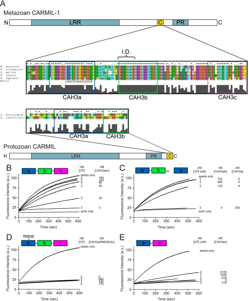
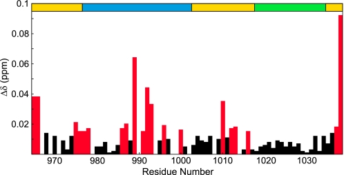
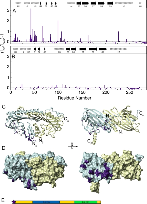
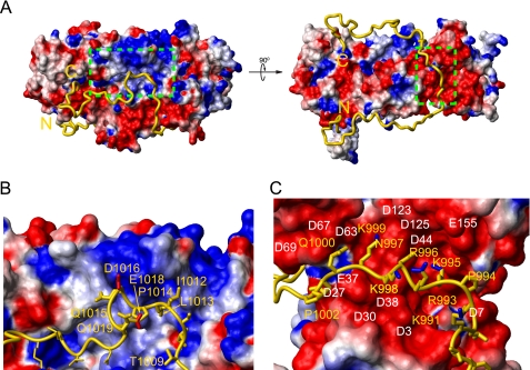
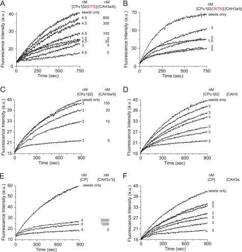
Capping protein (CP) is a ubiquitously expressed, 62-kDa heterodimer that binds the barbed end of the actin filament with approximately 0.1 nm affinity to prevent further monomer addition. CARMIL is a multidomain protein, present from protozoa to mammals, that binds CP and is important for normal actin dynamics in vivo. The CARMIL CP binding site resides in its CAH3 domain (CARMIL homology domain 3) located at or near the protein's C terminus. CAH3 binds CP with approximately 1 nm affinity, resulting in a complex with weak capping activity (30-200 nm). Solution assays and single-molecule imaging show that CAH3 binds CP already present on the barbed end, causing a 300-fold increase in the dissociation rate of CP from the end (i.e. uncapping). Here we used nuclear magnetic resonance (NMR) to define the molecular interaction between the minimal CAH3 domain (CAH3a/b) of mouse CARMIL-1 and CP. Specifically, we show that the highly basic CAH3a subdomain is required for the high affinity interaction of CAH3 with a complementary "acidic groove" on CP opposite its actin-binding surface. This CAH3a-CP interaction orients the CAH3b subdomain, which we show is also required for potent anti-CP activity, directly adjacent to the basic patch of CP, shown previously to be required for CP association to and high affinity interaction with the barbed end. The importance of specific residue interactions between CP and CAH3a/b was confirmed by site-directed mutagenesis of both proteins. Together, these results offer a mechanistic explanation for the barbed end uncapping activity of CARMIL, and they identify the basic patch on CP as a crucial regulatory site.


Figures

FIGURE 1.
FIGURE 1.
The organization of CARMIL proteins and the conserved CAH3 domain and the identification of CAH3a/b as the minimal CAH3 domain-containing fragment of mCARMIL-1. A, domain organization of protozoan and metazoan CARMIL proteins. Both contain a leucine-rich repeat domain (blue), a proline-rich (PR) domain (blue), and a CAH3 domain. The CAH3 domain (gold), which is necessary and sufficient for the anti-CP activities of CARMIL, is N-terminal to the PR domain in metazoan CARMIL proteins and C-terminal to the PR domain in protozoan CARMIL proteins. Sequence alignments of the CAH3 domains from several species are also shown. These alignments reveal that metazoan CARMIL CAH3 domains can be subdivided into three conserved subdomains, CAH3a, CAH3b, and CAH3c, whereas protozoan CARMIL CAH3 domains contain only the CAH3a and CAH3b subdomains. The consensus CP-binding motif LXHXTXXRPKX6P (21) is indicated, as is the capping “interference domain” (I.D.) defined in this work. B–E, shown are actin polymerization assays using 10% pyrene-labeled G-actin, actin seeds, and the indicated concentrations of CP and the following CAH3 domain fragments of mCARMIL-1: CAH3a/b/c (B), CAH3a/b or CAH3a only (C), CAH3a(R993E)/b/c (D), or CAH3a/c (E). Actin polymerization was monitored by pyrene fluorescence. The CAH3a, CAH3b, and CAH3c subdomains are shaded green, pink, and blue, respectively.
FIGURE 2.
FIGURE 2.
Chemical shift map of CP binding on CAH3a/b. Shown are the chemical shift change magnitudes of residues in CAH3a/b, plotted versus residue number. Significant values (Δδ > 0.015 ppm) are highlighted red. The domain organization of CAH3a/b is shown at the top (gold), with the CAH3a subdomain (blue) and the CAH3b subdomain (green) highlighted.
FIGURE 3.
FIGURE 3.
Chemical shift map of CAH3a/b binding on CP. Shown are the chemical shift change magnitudes of residues in CPα (A) and CPβ (B), plotted versus residue number. Significant values (Δδ > 0.10 ppm) are highlighted orange. The secondary structures for each subunit are shown at the top.7 Residues experiencing chemical shift changes are highlighted on the crystal structure of CP (C), including residues qualitatively assigned as being affected by CAH3a/b binding.
FIGURE 4.
FIGURE 4.
Intermolecular paramagnetic relaxation enhancement from N-terminally labeled CAH3a/b to CP. For this sample, a short extension of GSWGC replaced the CARMIL CAH3a/b N terminus (Ser964) for labeling through an iodoacetamido-reactive spin label to the -SH group of Cys963. The intensity ratio of each CP resonance peak in its diamagnetic form compared with its paramagnetic form is plotted versus residue number for CPα (A) and CPβ (B). The secondary structures for each subunit are shown at the top, as in Fig. 2. The ribbon diagram in C, where the N and C termini of each subunit are indicated, shows the orientation of the surface diagram in D and of those in supplemental Figs. S4-S7. CP subunits are shaded the same as in Fig. 2. Residues experiencing significant PRE (Id/Ip > 1.7) are highlighted on CP in D in purple. The location of the paramagnetic label is shown as a star on the domain diagram of CAH3a/b (E), where the coloring is the same as in Fig. 2.
FIGURE 5.
FIGURE 5.
The structure of the CP·CAH3a/b complex. CP is displayed as a surface charge diagram (A) in the same orientation as in Fig. 3. CAH3a/b (gold) is shown as a ribbon with its N and C termini indicated. The green boxed area on the left in A indicates the basic patch on CP and is shown enlarged in C. The boxed area on the right in A represents the acidic groove on CP and is shown enlarged in B. Selected residues from CAH3a/b (yellow) and CPβ (white) are indicated in B and C, and the side chains of CAH3a/b residues are shown as gold sticks with side chain C–NH bonds highlighted blue in B and side chain C–O bonds highlighted red in C.
FIGURE 6.
FIGURE 6.
Site-directed mutagenesis of CP and CAH3a/b. Pyrene-actin polymerization assays were performed as in Fig. 1. The concentrations of CP and CAH3a/b are indicated for each experiment. Two CP mutants were tested for their responsiveness to CAH3a/b (A and B). CAH3a/b mutants were tested for their ability to rescue actin polymerization (C–F). CAH3a/b (A–C) corresponds to residues Ser964–Val1038 of mCARMIL-1, CAH3a (D) corresponds to residues Ser964–Gln1019, CAH3a*b (E) contains the R993E mutation, and CAH3ab* (F) contains Val1026, Val1030, Phe1033, and Phe1034 mutations. a.u., absorbance units.

References

    1. Wear M. A., Cooper J. A. (2004) Trends Biochem. Sci. 29, 418–428 - PubMed
    1. Cooper J. A., Sept D. (2008) Int. Rev. Cell Mol. Biol. 267, 183–206 - PMC - PubMed
    1. Loisel T. P., Boujemaa R., Pantaloni D., Carlier M. F. (1999) Nature 401, 613–616 - PubMed
    1. Iwasa J. H., Mullins R. D. (2007) Curr. Biol. 17, 395–406 - PMC - PubMed
    1. Akin O., Mullins R. D. (2008) Cell 133, 841–851 - PMC - PubMed

Publication types

Associated data

LinkOut - more resources