. 2010 Aug;137(16):2785-94.
doi: 10.1242/dev.045997. Epub 2010 Jul 14.

Macroscopic stiffening of embryonic tissues via microtubules, RhoGEF and the assembly of contractile bundles of actomyosin

Affiliations

Macroscopic stiffening of embryonic tissues via microtubules, RhoGEF and the assembly of contractile bundles of actomyosin

Jian Zhou et al. Development. 2010 Aug.

Abstract

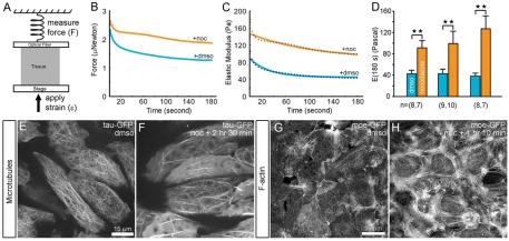
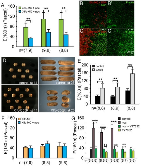
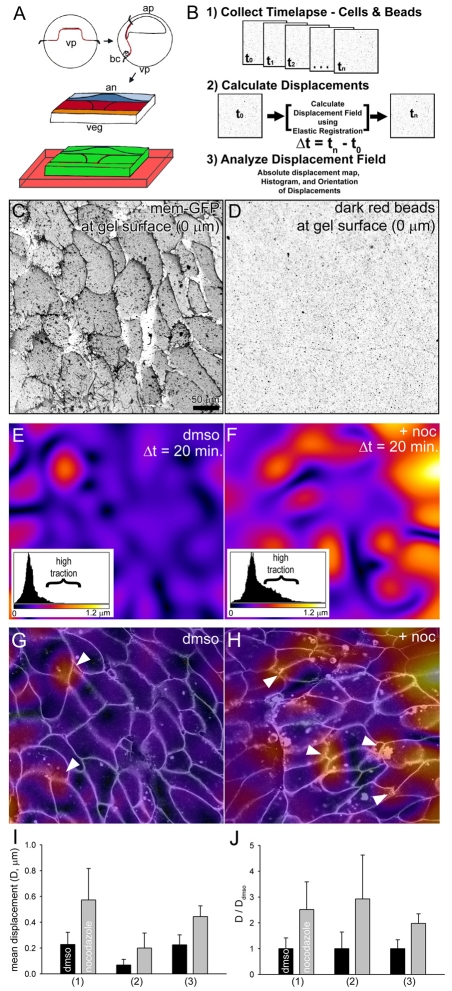
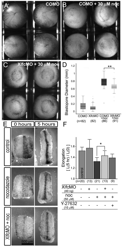
During morphogenesis, forces generated by cells are coordinated and channeled by the viscoelastic properties of the embryo. Microtubules and F-actin are considered to be two of the most important structural elements within living cells accounting for both force production and mechanical stiffness. In this paper, we investigate the contribution of microtubules to the stiffness of converging and extending dorsal tissues in Xenopus laevis embryos using cell biological, biophysical and embryological techniques. Surprisingly, we discovered that depolymerizing microtubules stiffens embryonic tissues by three- to fourfold. We attribute tissue stiffening to Xlfc, a previously identified RhoGEF, which binds microtubules and regulates the actomyosin cytoskeleton. Combining drug treatments and Xlfc activation and knockdown lead us to the conclusion that mechanical properties of tissues such as viscoelasticity can be regulated through RhoGTPase pathways and rule out a direct contribution of microtubules to tissue stiffness in the frog embryo. We can rescue nocodazole-induced stiffening with drugs that reduce actomyosin contractility and can partially rescue morphogenetic defects that affect stiffened embryos. We support these conclusions with a multi-scale analysis of cytoskeletal dynamics, tissue-scale traction and measurements of tissue stiffness to separate the role of microtubules from RhoGEF activation. These findings suggest a re-evaluation of the effects of nocodazole and increased focus on the role of Rho family GTPases as regulators of the mechanical properties of cells and their mechanical interactions with surrounding tissues.


Figures

Fig. 1.
Fig. 1.
Nocodazole stiffens embryonic tissues. (A) The elastic stiffness of early neural groove stage dorsal isolates (stage 16) are measured using a uniaxial unconfined compression test. (B) Representative plots of forces generated by two explants as they resist compression beginning at 0 seconds (blue line indicates explant incubated in DMSO; orange line indicates explant culture 40 minutes in 50 μM nocodazole). (C) Representative time-dependent elastic modulus of the two explants are calculated from the forces measured in B, the initial strain imposed on each isolate, and the transverse cross-sectional area of each isolate. Broken lines indicate the viscoelastic standard linear solid model fitted to the time-dependent modulus. (D) Residual elastic or Young's modulus, E(180), of 7 to 10 samples tested over three clutches or cohorts show nocodazole induces highly significant stiffening of dorsal isolates. We use the term `stiffness' for convenience when referring to E(180s). Double asterisks indicate highly significant differences (P<0.01). (E) Tau-GFP expressed in mesodermal cells in marginal zone explants reports the presence of microtubules and (F) that nocodazole reduces the amount of microtubules. (G,H) Moe-GFP expressed in mesodermal cells in marginal zone explants reports (G) the presence of F-actin and (H) that nocodazole-treatment leads to increased amounts of F-actin over the same timescale.
Fig. 2.
Fig. 2.
Xlfc is both necessary and sufficient to stiffen dorsal isolates and induce F-actin assembly, and acts in part through Rho kinase. (A) Xlfc knock-down (Xlfc-MO) reduces nocodazole-induced stiffening of dorsal isolates. (B) Embryos with one blastomere injected at the four-cell stage with Xlfc-MO and rhodamine dextran amine (RDA) show no changes in endogenous F-actin levels. (B′) F-actin levels alone in same field as in B. (C) Endogenous F-actin levels increase in the uninjected side once embryos are incubated with 50 μM nocodazole. (B′ and C′ show levels of F-actin without RDA-labeled channels of B and C, respectively.) (D) Embryos expressing low quantities of the constitutively activated Xflc point-mutant Xflc-C55R close their blastopore but form dorsally shortened embryos. (E) Dorsal isolates from Xlfc-C55R-expressing embryos are twofold stiffer than controls. (F) Microtubules do not contribute to stiffness. Xlfc-MO injected embryos treated with and without nocodazole show that microtubules do not contribute directly to stiffness. (G) Nocodazole-induced stiffening is reduced by the Rho kinase inhibitor Y27632. Stiffness measurements of dorsal isolates incubated in DMSO carrier, 50 μM nocodazole, and nocodazole and 40 μM Y27632 show stiffness is restored to normal control levels after nocodazole and Y27632 are combined (P<0.01; one clutch). Stiffness of isolates from three clutches show stiffness of nocodazole-treated tissues are significantly reduced after Y27632 treatment. Additional trials from four clutches treated with both 50 μM nocodazole and 40 μM Y27632 are also significantly stiffer than those treated with Y27632 alone. Comparisons of dorsal isolate stiffness within each clutch are tested for significance using the Mann-Whitney U-test and significance of stiffness measurements among multiple clutches were calculated using one-way ANOVA (**P<0.01; ***P<0.005).
Fig. 3.
Fig. 3.
Traction maps reveals nocodazole increases physical contractility of tissue. (A) Assembly of marginal zone explants on displacement-reporting polyacrylamide gels. (B) Traction maps are calculated from confocal time-lapse sequences of gel, cell and tissue movements. (C) Confocal section of mesodermal cells expressing a mem-GFP cultured on fibronectin-conjugated polyacrylamide gel. (D) Confocal section collected at the same level shows the position of dark-red fluorescent microsphere beads on the surface of the gel. (E) Traction map shows the magnitude of bead displacements beneath mesodermal cells within an intact marginal zone explant incubated in DMSO. The inset histogram shows the frequency of bead displacements and the scale of displacements. Most displacements are limited to less than 0.3 μm within the color range of purple to blue; regions of high traction above 0.3 μm are within the color ranges of red to yellow. (F) Traction map of mesodermal cells in explants incubated in 50 μM nocodazole show large displacements spread over larger areas. The histogram shows a large increase in regions of high traction. (G,H) Combined displacement maps shown in E and F, respectively, with overlying cells outlines collected from a confocal section 5 μm deeper into the cells. High regions of traction in both DMSO and after incubation in nocodazole appear to colocalize with cell-cell junctions at the mediolateral ends of cells (arrowheads). (I) Mean displacements calculated from the full field of view collected from three explants from three clutches show significant and large increases in traction after explants are incubated in nocodazole. Variances from clutch to clutch may reflect slight changes in substrate preparation or clutch-to-clutch differences in cell-generated traction. (J) Mean displacements normalized for each clutch show nocodazole consistently increases traction in all cases.
Fig. 4.
Fig. 4.
Mechanical rescue of developmental defects. (A-D) Xlfc-MO produces a small but significant rescue of blastopore closure defects induced by nocodazole. (E,F) Xlfc-MO produces a moderate rescue of dorsal isolate elongation rates. (A) Embryos injected with control morpholinos close their blastopore. (B) Control morpholino-injected embryos cultured from stage 10.25 onwards in 30 μM nocodazole do not close their blastopore. (C) Embryos injected with Xlfc MO show some improvements in closing their blastopore. (D) Measurements of the diameter of the open-blastopore at stage 12.75 (just prior to closure in control embryos) show Xlfc MO provides a small but highly significant rescue of the nocodazole-induced open blastopore defect. (E) Dorsal isolates cultured from stage 13 onwards extend by nearly 50%. Incubation in 50 μM nocodazole reduces the degree of elongation, but prior injection with Xlfc MO can ameliorate that reduction. (F) Quantitative measurements of explants from three different clutches show that prior injection with XlfcMO can partly rescue the nocodazole-induced effects. By contrast, low doses of Y27632 do not provide significant rescue.

References

    1. Arganda-Carreras I., Sorzano C. O. S., Marabini R., Carazo J. M., de Solorzano C. O., Kybic J. (2006). Consistent and elastic registration of histological sections using vector-spline regularization. CVAMIA:Computer Vision Approaches to Medical Image Analysis 4241, 85-95
    1. Bakal C. J., Finan D., LaRose J., Wells C. D., Gish G., Kulkarni S., DeSepulveda P., Wilde A., Rottapel R. (2005). The Rho GTP exchange factor Lfc promotes spindle assembly in early mitosis. Proc. Natl. Acad. Sci. USA 102, 9529-9534 - PMC - PubMed
    1. Barentin C., Sawada Y., Rieu J. P. (2006). An iterative method to calculate forces exerted by single cells and multicellular assemblies from the detection of deformations of flexible substrates. Eur. Biophys. J. 35, 328-339 - PubMed
    1. Beningo K. A., Wang Y. L. (2002). Flexible substrata for the detection of cellular traction forces. Trends Cell. Biol. 12, 79-84 - PubMed
    1. Beningo K. A., Lo C. M., Wang Y. L. (2002). Flexible polyacrylamide substrata for the analysis of mechanical interactions at cell-substratum adhesions. Methods Cell Biol. 69, 325-339 - PubMed

Publication types

MeSH terms

Substances