. 2010 Nov;38(21):7570-8.
doi: 10.1093/nar/gkq620. Epub 2010 Jul 14.

A model in vitro system for co-transcriptional splicing

Affiliations

A model in vitro system for co-transcriptional splicing

Yong Yu et al. Nucleic Acids Res. 2010 Nov.

Abstract

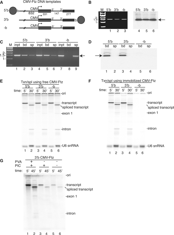
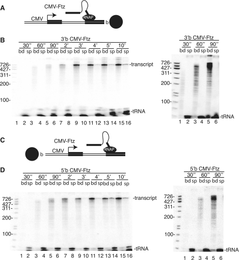
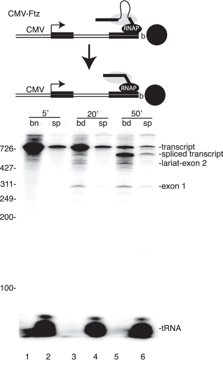
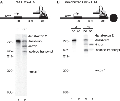
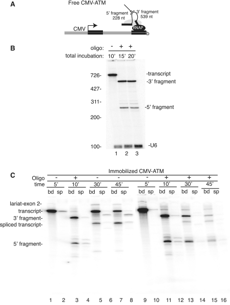
A hallmark of metazoan RNA polymerase II transcripts is the presence of numerous small exons surrounded by large introns. Abundant evidence indicates that splicing to excise introns occurs co-transcriptionally, prior to release of the nascent transcript from RNAP II. Here, we established an efficient model system for co-transcriptional splicing in vitro. In this system, CMV-DNA constructs immobilized on beads generate RNAP II transcripts containing two exons and an intron. Consistent with previous work, our data indicate that elongating nascent transcripts are tethered to RNAP II on the immobilized DNA template. We show that nascent transcripts that reach full length, but are still attached to RNAP II, are efficiently spliced. When the nascent transcript is cleaved within the intron using RNase H, both the 5' and 3' cleavage fragments are detected in the bound fraction, where they undergo splicing. Together, our work establishes a system for co-transcriptional splicing in vitro, in which the spliceosome containing the 5' and 3' exons are tethered to RNAP II for splicing.


Figures

Figure 1.
Figure 1.
Efficient RNAP II transcription and splicing on immobilized CMV-Ftz DNA templates. (A) Schematic of CMV-Ftz DNA templates are shown, indicating the position of the biotin. The sizes of the CMV promoter and Ftz exons and intron are indicated. The gray circle represents the streptavidin magnetic bead. (B) CMV-Ftz DNA templates containing biotin at the 5′ (5′b) or 3′ end (3′b), or no biotin (-b) were constructed by PCR and detected on an ethidium bromide gel (lanes 1–3) or by uniform labeling (lanes 4–6). (C) Immobilized 5′b, 3′b and –b CMV-Ftz DNA templates were mixed with streptavidin magnetic beads, and after washing, bound (bnd) and supernatant (sup) fractions were analyzed on a 1% agarose gel stained with ethidium bromide. Total input is shown (lanes 1, 4 and 7). (D) Biotinylated CMV-Ftz DNA templates were uniformly labeled, bound to streptavidin magnetic beads, followed by incubation in nuclear extract at 30° for 30 min. Bound (1, 3 and 5) and supernatant (2, 4 and 6) fractions were analyzed on a 6.5% denaturing polyacrylamide gel. The arrow in panels B, C and D indicate the Ftz DNA template. Marker in panels B and C are in kilobases. (E and F) Free (E) or immobilized (F) CMV-Ftz DNA templates were incubated under RNAP II transcription/splicing conditions for 5 min, actinomycin D was added, and incubation was continued for another 25 min. Total RNA was fractionated on a 6.5% denaturing polyacrylamide gel. The splicing intermediates and products, tRNA, and U6 snRNA are indicated. Ori designates the gel origin. (G) PIC conditions with or without PVA were compared with standard RNAP II transcription/splicing conditions. Total bound (lane 1, 3 and 5) and supernatant (lane 2, 4, 6) fractions were analyzed on a 6.5% denaturing polyacrylamide gel.
Figure 2.
Figure 2.
Transcription time course to analyze association of nascent transcripts with RNAP II bound to the DNA template. (A and C) Schematics showing transcription of 3′b (A) or 5′b (C) CMV-Ftz DNA template. (B and D) Immobilized 3′b (B) or 5′b (D) CMV-Ftz DNA template was incubated under PIC conditions with PVA for 30″, 60″, 90″, 2′, 3′, 4′, 5′and 10′. Total bound and supernatant fractions were separated, and the bound fractions were washed in 100 mM KCl, 0.1% triton. Bound (lanes 1, 3, 5, 7, 9 11, 13 and 15) and supernatant (lanes 2, 4, 6, 8, 10 12, 14 and 16) fractions were analyzed on a 6.5% denaturing polyacrylamide gel. (B, D) right. Longer exposure of lanes 1–6 for (B) and (D).
Figure 3.
Figure 3.
Evidence that nascent transcripts are spliced while tethered to RNAP II. Immobilized 3′b CMV-Ftz DNA template was incubated under PIC conditions with PVA for 5 min, actinomycin D was added, and incubation was continued for a total of 20 and 50 min. The bound and supernatant fractions were separated, and the bound fractions were washed in 100 mM KCl, 0.1% triton. Total RNA was isolated from the bound and supernatant fractions and run on a 6.5% denaturing polyacrylamide gel.
Figure 4.
Figure 4.
Splicing of free and immobilized CMV-ATM DNA template. (A) Free CMV-ATM DNA template was incubated under RNAP II transcription/splicing conditions for the times indicated and analyzed on a 6% denaturing polyacrylamide gel. The unspliced transcript, splicing intermediates and products are indicated. (B) 3′b CMV-ATM DNA template was immobilized on streptavidin magnetic beads and then incubated under PIC conditions with PVA. Total bound (lanes 1 and 3) and supernatant (lanes 2 and 4) fractions were analyzed on a 6% denaturing polyacrylamide gel.
Figure 5.
Figure 5.
The 5′ and 3′ portions of the transcript cleaved within the intron by RNase H are tethered to RNAP II and undergo splicing. (A) Schematic showing 5′ and 3′ portions of the ATM transcript cleaved within the intron. The short line indicates the oligonucleotide used for RNase H cleavage, and the sizes of the 5′ and 3′ cleavage products are indicated. (B) 3′b CMV-ATM DNA template was incubated under RNAP II transcription/splicing conditions for 10 min (lanes 1). The oligonucleotide was then added and incubation was continued for 5 (lane 2) or 10 min (lane 3). The RNA was cleaved by the endogenous RNase H in the nuclear extract, and the 5′ and 3′ cleavage fragments are indicated. (C) 3′b CMV-ATM DNA template was immobilized on streptavidin magnetic beads and then incubated under PIC conditions with PVA for 5 min (lanes 1 and 2) followed by addition of actinomycin D and oligonucleotide for 5 min (lanes 3 and 4), or only actinomycin D for 25 min (lanes 5 and 6) and 40 min (lanes 7 and 8). Lanes 9–12 are the same as lanes 1–4. An aliquot of the sample shown in lane 11 was incubated further, for a total of 30 and 45 min, and then RNA from total bound and supernatant fractions was analyzed on a 5.75% denaturing polyacrylamide gel (lanes 13–16).

Similar articles

Cited by

References

    1. Black DL. Mechanisms of alternative pre-messenger RNA splicing. Annu. Rev. Biochem. 2003;72:291–336. - PubMed
    1. Kornblihtt AR, de la Mata M, Fededa JP, Munoz MJ, Nogues G. Multiple links between transcription and splicing. RNA. 2004;10:1489–1498. - PMC - PubMed
    1. Pandit S, Wang D, Fu XD. Functional integration of transcriptional and RNA processing machineries. Curr. Opin. Cell Biol. 2008;20:260–265. - PMC - PubMed
    1. Wang ET, Sandberg R, Luo S, Khrebtukova I, Zhang L, Mayr C, Kingsmore SF, Schroth GP, Burge CB. Alternative isoform regulation in human tissue transcriptomes. Nature. 2008;456:470–476. - PMC - PubMed
    1. Nilsen TW, Graveley BR. Expansion of the eukaryotic proteome by alternative splicing. Nature. 2010;463:457–463. - PMC - PubMed

Publication types