. 2010 Aug 1;70(15):6139-49.
doi: 10.1158/0008-5472.CAN-10-0706. Epub 2010 Jul 14.

Gr-1+CD11b+ myeloid cells tip the balance of immune protection to tumor promotion in the premetastatic lung

Affiliations

Gr-1+CD11b+ myeloid cells tip the balance of immune protection to tumor promotion in the premetastatic lung

Hannah H Yan et al. Cancer Res. .

Abstract

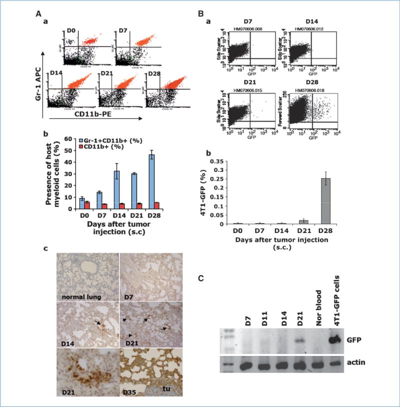
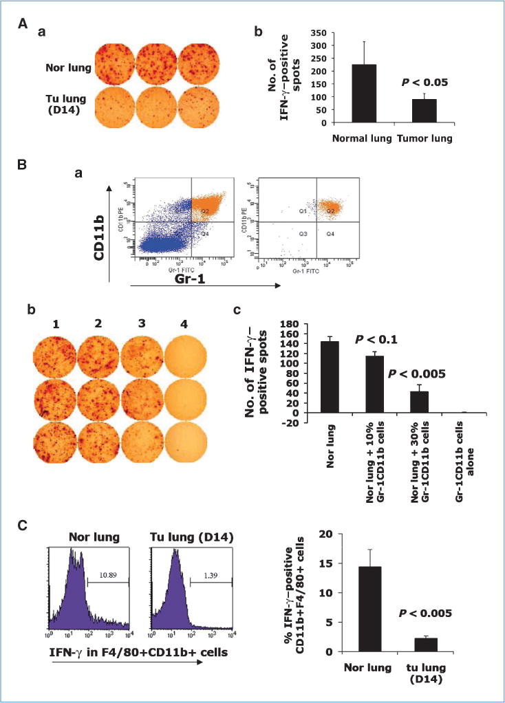
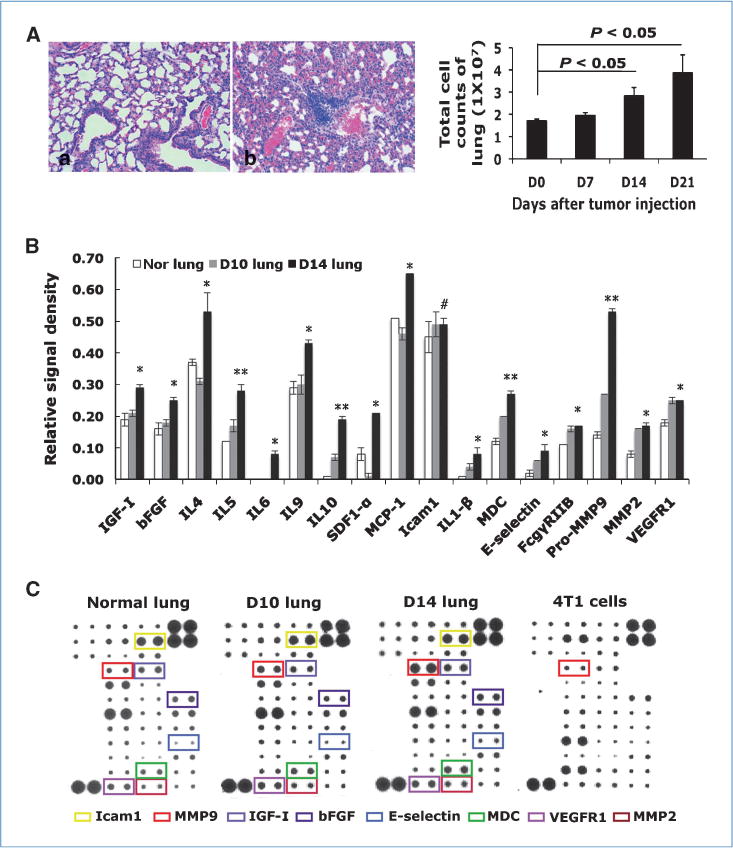
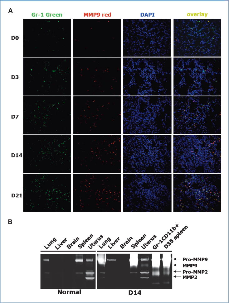
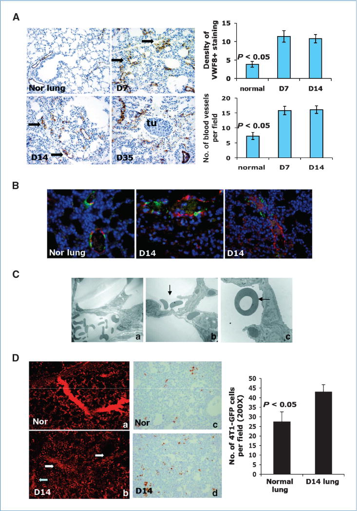
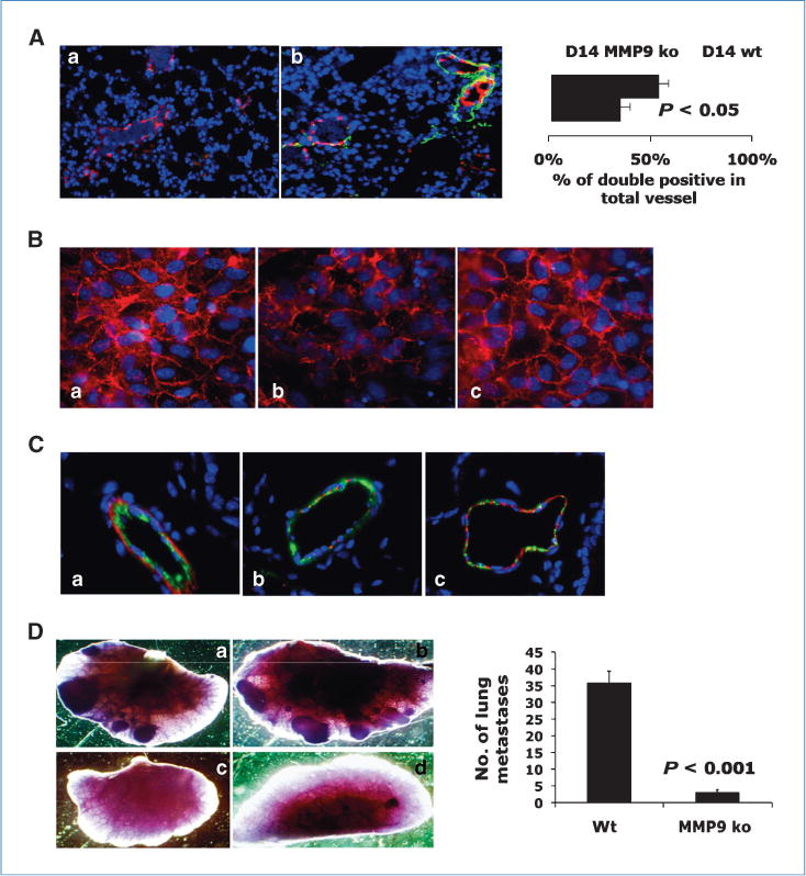
The mechanisms by which a primary tumor affects a selected distant organ before tumor cell arrival remain to be elucidated. This report shows that Gr-1+CD11b+ cells are significantly increased in lungs of mice bearing mammary adenocarcinomas before tumor cell arrival. In the premetastatic lungs, these immature myeloid cells significantly decrease IFN-gamma production and increase proinflammatory cytokines. In addition, they produce large quantities of matrix metalloproteinase 9 (MMP9) and promote vascular remodeling. Deletion of MMP9 normalizes aberrant vasculature in the premetastatic lung and diminishes lung metastasis. The production and activity of MMP9 is selectively restricted to lungs and organs with a large number of Gr-1+CD11b+ cells. Our work reveals a novel protumor mechanism for Gr-1+CD11b+ cells that changes the premetastatic lung into an inflammatory and proliferative environment, diminishes immune protection, and promotes metastasis through aberrant vasculature formation. Thus, inhibition of Gr-1+CD11b+ cells could normalize the premetastatic lung environment, improve host immunosurveillance, and inhibit tumor metastasis.


Conflict of interest statement

Disclosure of Potential Conflicts of Interest

No potential conflicts of interest were disclosed.

Figures

Figure 1
Figure 1
Gr-1+CD11b+ cells infiltrate into the lungs of mice bearing 4T1 tumors prior to tumor cell arrival. D0, non–tumor-bearing mice; D7, D14, D21, and D35, days after 4T1 tumor inoculation (s.c.). Aa, flow cytometry of Gr-1+CD11b+ cells in total lung single-cell suspension. Ab, quantitative data for Aa. Ac, IHC of Gr-1+CD11b+ cells in lung sections. Bottom left, enlarged Gr-1+CD11b+ cell cluster. Tu, a tumor metastasis nodule. Ba, flow cytometry of 4T1-GFP tumor cells in lungs. Bb, quantitative data for Ba. C, GFP-PCR in nucleated peripheral blood cells from mice bearing 4T1 tumors. Shown is one of the two experiments performed. Three to four mice per group were examined for all experiments.
Figure 2
Figure 2
Gr-1+CD11b+ cells inhibit IFN-γ production in premetastatic lung. Aa, IFN-γ ELISPOT from normal lungs and day 14 lungs. Ab, quantitative data from Aa. Ba, Gr-1+CD11b+ cells before (left) and after (right) sorting. Bb, IFN-γ ELISPOT of normal lung single-cell suspension cocultured with sorted Gr-1+CD11b+ cells. Column 1, normal lung alone (2 × 10⁶); columns 2 to 3, coculture of normal lung (2 × 10⁶) with Gr-1+CD11b+ cells (2 × 10⁵ for column 2, 1 × 10⁶ for column 3); column 4, Gr-1+CD11b+ cells alone (2 × 10⁶). Bc, quantitative results for Bb. Samples are in triplicate. One experiment of two is shown. C, flow cytometry analysis of IFN-γ expression in F4/80 +CD11b+ cells in lungs of normal and 4T1 tumor-bearing mice. Three to four mice were analyzed; right, the quantitative data.
Figure 3
Figure 3
Cytokine/chemokine profiling of premetastatic lung and normal lung. A, increased tissue density in premetastatic lung by H&E staining (Aa, normal lung; Ab, day 14 lung) and total cell counts from lungs at different days after tumor injection (right). Three to four mice per group. B, relative signal intensity from cytokine/chemokine array. *, P < 0.05; **, P < 0.01; #, P > 0.05. C, cytokine/chemokine array. Cytokine of interest is labeled with a specific color. Shown is one of the two experiments performed.
Figure 4
Figure 4
Significantly elevated MMP9 in premetastatic lung with Gr-1+CD11b+ cells as the major resource. A, co-IF staining of Gr-1 (green) and MMP9 (red) in lungs at different day after tumor injection. Blue, nuclei. One experiment from three is shown. B, gelatin zymography of protein lysis from indicated organs of normal mice and mice bearing 14 day 4T1 tumors. One of three experiments is shown.
Figure 5
Figure 5
Aberrant and leaky vasculature in premetastatic lung. A, VWF8 staining. Days after s.c. tumor inoculation are indicated. Arrows, vessel-like structure. Tu, metastasis nodules. Quantitative data were obtained in a blinded fashion (top right and bottom). B, co-IF staining of VWF8 (red) and αSMA (green). C, electron microscopy of blood vessel in normal lung (Ca) and day 14 lung (Cb–c). Arrows, RBC in alveoli. Two to three mice were examined. D, vascular leakage in premetastatic lung; Da and b, confocal imaging. Four to five mice were examined. Dc and d, IHC of GFP to detect 4T1-GFP tumor cells in the lungs. Three to four mice were examined. GFP-positive cells were counted in a blinded fashion (right).
Figure 6
Figure 6
Deletion of MMP9 in host decreased aberrant vascular phenotype and diminished lung metastasis. A, co-IF staining of VWF8 (red) and αSMA (green) in lungs of wt or MMP9 ko bearing 4T1 tumors day 14 after injection. Right, quantitative data. Three to four mice examined. B, VE-cadherin staining (red) of HUVEC cocultured with wt (b) or MMP9 ko Gr-1+CD11b+ cells (c), or HUVEC alone (a). One of two experiments is shown. C, co-IF staining of VWF8 (green) and VE-cadherin (red) in lungs of nontumor-bearing (a), wt (b), and MMP9 ko mice (c) bearing 4T1 tumors day 14 after injection. D, whole-lung mounts of wt (a and b) and MMP9 ko (c and d) mice bearing 4T1 tumors 35 d after s.c. injection. Right, number of lung metastatic nodules (P < 0.001). Five to six mice per group.

References

    1. Steeg PS. Tumor metastasis: mechanistic insights and clinical challenges. Nat Med. 2006;12:895–904. - PubMed
    1. Hynes RO. Metastatic potential: generic predisposition of the primary tumor or rare, metastatic variants-or both? Cell. 2003;113:821–3. - PubMed
    1. Kang Y, Siegel PM, Shu W, et al. A multigenic program mediating breast cancer metastasis to bone. Cancer Cell. 2003;3:537–49. - PubMed
    1. Minn AJ, Gupta GP, Siegel PM, et al. Genes that mediate breast cancer metastasis to lung. Nature. 2005;436:518–24. - PMC - PubMed
    1. Gupta GP, Massague J. Cancer metastasis: building a framework. Cell. 2006;127:679–95. - PubMed

Publication types

MeSH terms