. 2010 Oct;84(19):9897-906.
doi: 10.1128/JVI.00761-10. Epub 2010 Jul 14.

Two overlapping domains of a lyssavirus matrix protein that acts on different cell death pathways

Affiliations

Two overlapping domains of a lyssavirus matrix protein that acts on different cell death pathways

Florence Larrous et al. J Virol. 2010 Oct.

Abstract

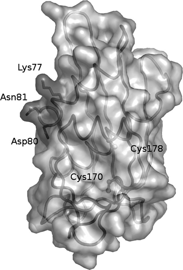
The lyssavirus matrix (M) protein induces apoptosis. The regions of the M protein that are essential for triggering cell death pathways are not yet clearly defined. We therefore compared the M proteins from two viruses that have contrasting characteristics in terms of cellular apoptosis: a genotype 3 lyssavirus, Mokola virus (MOK), and a genotype 1 rabies virus isolated from a dog from Thailand (THA). We identified a 20-amino-acid fragment (corresponding to positions 67 to 86) that retained the cell death activities of the full-length M protein from MOK via both the tumor necrosis factor-related apoptosis-inducing ligand (TRAIL) and inhibition of cytochrome c oxidase (CcO) activity. We found that the amino acids at positions 77 and 81 have an essential role in triggering these two cell death pathways. Directed mutagenesis demonstrated that the amino acid at position 77 affects CcO activity, whereas the amino acid at position 81 affects TRAIL-dependent apoptosis. Mutations in the full-length M protein that compromised induction of either of these two pathways resulted in delayed apoptosis compared with the time to apoptosis for the nonmutated control.


Figures

FIG. 1.
FIG. 1.
M1-7-induced apoptosis by the extrinsic and intrinsic pathways. (A) Schematic representation of M-MOK mutants with deletions N-terminally fused to an EGFP protein tag. The α helix (light gray) and β sheets (dark gray) were determined by the study of M-LAG (15). HeLa cells were transfected with plasmids encoding EGFP alone (eGFP), full-length M (M), or truncated forms (M1, M2, M1-1, M1-2, M1-5, M1-6, and M1-7). Cells were harvested after 24 h of incubation at 37°C. (B) The percentage of transfected cells undergoing apoptosis was measured by TUNEL assay. Transfections were performed in the presence or absence of a neutralizing anti-TRAIL antibody (1 μg of anti-TRAIL for 104 cells); this antibody inhibits TRAIL-mediated apoptosis. (C) HeLa cells were transfected in the presence of soluble TRAIL receptors R1 and R2. Their effect on apoptosis was measured by TUNEL assay. (D) HeLa cells were transfected with the various mutant constructs and cultured; the supernatants were collected, clarified, and added to freshly prepared cell cultures. The cultures were incubated at 37°C for 24 h in the presence or absence of anti-TRAIL antibody (1 μg of anti-TRAIL for 104 cells). The percentage of cells undergoing apoptosis was measured by TUNEL assay. The results are expressed as the mean value for three independent experiments. Error bars indicate standard deviations of these values. Significant differences before and after treatment with anti-TRAIL antibody or R1 and R2 receptors are indicated by asterisks (P < 0.05). (E) CcO activity was evaluated after 24 h. The reported values are the percentages of CcO activity relative to that of cells expressing EGFP alone normalized to the protein concentration. Mean values for three independent experiments and error bars are shown. Significant differences between cells expressing EGFP and the mutants are indicated by asterisks (P < 0.05).
FIG. 2.
FIG. 2.
Localization of M-truncated mutants and M1-7 mutants in the transfected cells. (A) Mitochondrion-enriched fractions (mito) and cytosolic fractions (cyto) were analyzed by Western blotting and densitometry. For each mutant, the ratio of the signal in the mitochondrion-enriched fraction to that in the cytosolic fraction was determined, and the mean for two independent experiments is reported. (B) Representative immunoblot with anti-EGFP antibody: different times of exposure to the film are used to show EGFP-tagged constructs. However, CcO4 and β-actin bands are obtained from single exposure times. (C) HeLa cells were cotransfected with plasmid vectors expressing DsRed2-Mito and constructs encoding mutants with deletions (M1-2, M1-5, M1-6, M1-7) or the vector alone (EGFP). The subcellular localization of these constructs was assessed after 24 h with a Zeiss Axioplan 2.2 fluorescence microscope equipped with a Zeiss ApoTome system. EGFP, the EGFP signal, DsRed2 (DsRed2-Mito), the mitochondrial marker; Merge, overlay image composed of the signals for various constructs and the mitochondrial marker.
FIG. 3.
FIG. 3.
Role of positions K77 and N81 and the α1 helix of the M1-7 fragment in apoptosis pathways. (A) Sequence alignment of sequence positions 67 to 86 of lyssavirus matrix protein. The amino acids at positions 77 and 81 are indicated in boldface. (B) Schematic representation of M1-7 mutants with deletions N-terminally fused to an EGFP protein tag. Mutations were introduced into each subfragment at position 77 or 81. M1-8 was also mutated at position 74 (M1-8 P74). (C). HeLa cells were transfected with plasmids expressing EGFP alone (eGFP), M1-7 KN (wild type for MOK sequence) or its mutated forms (M1-7 EN, KE, and RE), or truncated mutants M1-8 K and its E77 (M1-8 E), R77 (M1-8 R), and P74 (M1-P74) mutated forms and M1-9 and its E81 mutated form (M1-9 E). The transfected cells were cultured in the presence or in the absence of a neutralizing anti-TRAIL antibody (1 μg of anti-TRAIL). After 24 h of incubation at 37°C the percentage of transfected cells undergoing apoptosis was measured by TUNEL assay. The results are expressed as mean values for three independent experiments. Error bars indicate standard deviations of these values. Significant differences in the presence and absence of treatment with anti-TRAIL antibody are indicated with asterisks (P < 0.05). (D) CcO activity was assayed after 24 h of culture. Reported results are percentages of activity relative to that of cells expressing EGFP alone (arbitrarily defined to be 100%) and normalized for protein concentration. Values are means for three independent experiments, and standard deviations are shown as error bars. Significant differences between cells expressing EGFP and the mutants are indicated by asterisks (P < 0.05). (E) HeLa cells were cotransfected with plasmid vectors encoding DsRed2-Mito and plasmids encoding mutants M1-7 EN, KE, and RE and mutants M1-8 and M1-9 or their mutants with point mutations (M1-8 E, M1-8 R, and M1-9 E). The subcellular localization of these mutants was assessed after 24 h with a Zeiss Axioplan 2.2 fluorescence microscope equipped with a Zeiss ApoTome system. eGFP, EGFP signal, DsRed2 (DsRed2-Mito), mitochondrial marker; Merge, overlay image between the various constructs and the mitochondrial pattern of the cell.
FIG. 4.
FIG. 4.
Pivotal role of positions 77 and 81 of M protein on the induction of apopotosis by the intrinsic and extrinsic pathways. M-MOK KN was mutated by site-directed mutagenesis to M-MOK RE, and M-THA RE was mutated to M-THA KN. HeLa cells were transfected with the constructs encoding these mutants and with the vector alone. (A) The transfected cells were cultured in the presence or absence of a neutralizing anti-TRAIL antibody (1 μg of anti-TRAIL) for 24 and 48 h, and the percentage of transfected cells undergoing apoptosis was measured by TUNEL assay. (B) The CcO activity was measured after 24 h of culture. CcO activity is reported as a percentage of that in cells expressing eGFP alone and normalized to the protein concentration. Values are means for three independent experiments, and error bars indicate the standard deviations. Significant differences between cells expressing EGFP and the mutants are indicated with asterisks (P < 0.05). (C) Cytosol and mitochondrion-enriched fractions were tested for the presence of the mutants by Western blotting and densitometry, and the results reported are the ratio of the EGFP signal in the mitochondrion-enriched fraction to that in the cytosol fraction. (D) A representative immunoblot of fractions of cells expressing M-MOK and M-THA mutants probed with anti-EGFP antibody is shown. Different times of exposure to the film were used to reveal EGFP-tagged constructs. However, CcO-4 and β-actin were obtained from single exposure times.
FIG. 5.
FIG. 5.
Representation of the structure of the LAG matrix protein from residues 48 to 202 after a crystallography study (2). Positions K77 and N81 are at the outside face of the protein.

Similar articles

Cited by

References

    1. Ashkenazi, A., and R. S. Herbst. 2008. To kill a tumor cell: the potential of proapoptotic receptor agonists. J. Clin. Invest. 118:1979-1990. - PMC - PubMed
    1. Assenberg, R., O. Delmas, S. C. Graham, A. Verma, N. Berrow, D. I. Stuart, R. J. Owens, H. Bourhy, and J. M. Grimes. 2008. Expression, purification and crystallization of a lyssavirus matrix (M) protein. Acta Crystallogr. Sect. F Struct. Biol. Cryst. Commun. 64:258-262. - PMC - PubMed
    1. Badrane, H., C. Bahloul, P. Perrin, and N. Tordo. 2001. Evidence of two Lyssavirus phylogroups with distinct pathogenicity and immunogenicity. J. Virol. 75:3268-3276. - PMC - PubMed
    1. Baloul, L., and M. Lafon. 2003. Apoptosis and rabies virus neuroinvasion. Biochimie 85:777-788. - PubMed
    1. Benedict, C. A., P. S. Norris, and C. F. Ware. 2002. To kill or be killed: viral evasion of apoptosis. Nat. Immunol. 3:1013-1018. - PubMed

Publication types

MeSH terms

LinkOut - more resources