. 2010 Oct;84(19):10087-101.
doi: 10.1128/JVI.02425-09. Epub 2010 Jul 14.

Development of a targeted gene vector platform based on simian adenovirus serotype 24

Affiliations

Development of a targeted gene vector platform based on simian adenovirus serotype 24

Natalya Belousova et al. J Virol. 2010 Oct.

Abstract

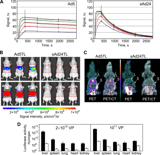
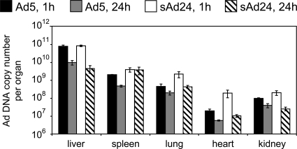
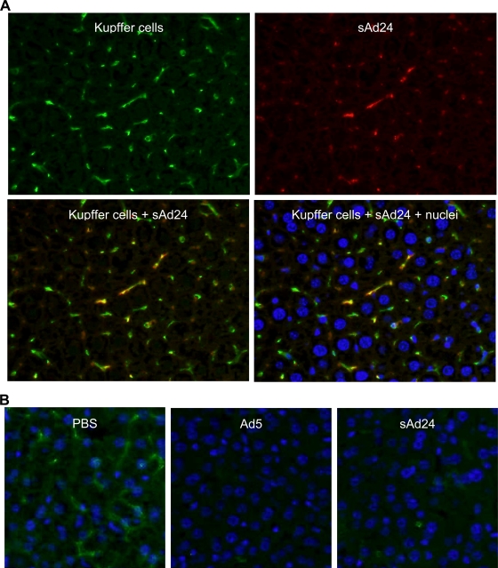
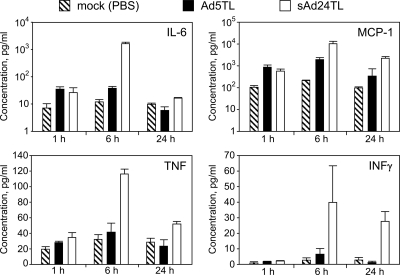
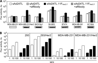
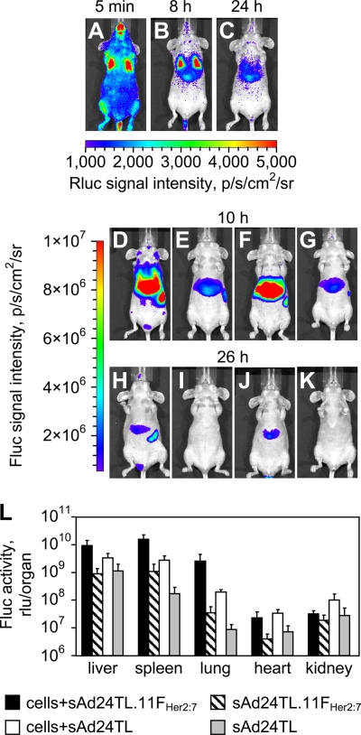
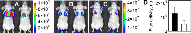
Efforts to develop adenovirus vectors suitable for genetic interventions in humans have identified three major limitations of the most frequently used vector prototype, human adenovirus serotype 5 (Ad5). These limitations--widespread preexisting anti-Ad5 immunity in humans, the high rate of transduction of normal nontarget tissues, and the lack of target-specific gene delivery--justify the exploration of other Ad serotypes as vector prototypes. In this paper, we describe the development of an alternative vector platform using simian Ad serotype 24 (sAd24). We found that sAd24 virions formed unstable complexes with blood coagulation factor X and, because of that, transduced the liver and other organs at low levels when administered intravenously. The overall pattern of biodistribution of sAd24 particles was similar, however, to that of Ad5, and the intravenously injected sAd24 was cleared by Kupffer cells, leading to their depletion. We modified the virus's fiber protein to design a Her2-specific derivative of sAd24 capable of infecting target human tumor cells in vitro. In the presence of neutralizing anti-Ad5 antibodies, Her2-mediated infection with targeted sAd24 compared favorably to that with the Ad5-derived vector. When used to target Her2-expressing tumors in animals, this fiber-modified vector achieved a higher level of gene transfer to metastasis-containing murine lungs than to tumor-free lungs. In aggregate, these studies provide important insights into sAd24 biology, identify its advantages and limitations as a vector prototype, and are thus essential for further development of an sAd24-based gene delivery platform.


Figures

FIG. 1.
FIG. 1.
Instability of FX-sAd24 complexes results in reduced liver transduction by sAd24-derived vector. (A) The interactions of Ad5 and sAd24 virions with FX were analyzed using surface plasmon resonance. Viral particles bound to the biosensor chip (Ad5 at a coating level of 1,387 resonance units [ru] and sAd24 at 1,699 ru) were probed with soluble FX. Colored lines correspond to the signals obtained in duplicate runs at each tested concentration of FX (blue, 12.5 nM; red, 25 nM; brown, 50 nM; green, 100 nM; and black, 200 nM). As evidenced by the dissociation part of the sensorgrams, the FX-sAd24 complexes were much less stable than the FX-Ad5 complexes. (B) Whole-body images of female nude mice that were injected intravenously (tail vein) with an Ad5- or an sAd24-derived vector, each expressing a dual-modality TL reporter (HSV tk fused with Fluc). Mice injected with 2 × 10¹⁰ VP (upper panels) and 1011 VP (lower panels) of either Ad5TL (left panels) or sAd24TL (right panels) are shown. The intensities of the light signals in the selected regions of interest (red squares) are shown as pseudocolor overlays. Here and in Fig. 8 and 9, the pseudocolor scale shows the signal intensity range. Notably, the relative infectivities of Ad5TL and sAd24TL, which had been determined by the spot assay in 293/Her2 cells in vitro, were 10 VP/infectious units and 53 VP/infectious units, respectively. p/s/cm2/sr, photons/s/cm2/steradian. (C) On the left on each panel, PET images (coronal slices) of Ad-injected animals enabled by 18F-labeled FEAU are shown. On the right, these PET images are superimposed on the CT images that provide anatomic landmarks. The white-dotted contours show the tracer uptake by the livers (L) and the spleens (S) of the animals. The signals in the bladders (B) are due to physiologic accumulation of the small-molecule tracer in the urine rather than vector-expressed HSV tk activity. The pseudocolor scale between the panels shows the PET signal intensity range, from 0 (black) to 6 times the mean muscle uptake value (white). (D) Activities of Fluc reporter in homogenates of tissue samples collected from mice injected with 2 × 10¹⁰ VP or 1011 VP of either Ad5TL (black bars) or sAd24TL (white bars) is shown. Each bar represents an average signal intensity (shown in relative light units [rlu] per entire organ) measured in organ samples obtained from five animals. Error bars indicate the standard deviations calculated for duplicate data points.
FIG. 2.
FIG. 2.
Ad5 and sAd24 virions show similar patterns of in vivo distribution. Shown are the copy numbers of Ad5 and sAd24 viral genomes per organ detected by qPCR in isolated murine organs at 1 h and 24 h after systemic administration of the vectors. Error bars indicate the standard deviations calculated for duplicate data points.
FIG. 3.
FIG. 3.
Uptake of sAd24 virions by KCs in the liver leads to KC depletion. (A) Immunofluorescence staining of the liver collected from a mouse injected with sAd24 vector 10 min after injection. The upper panels show the liver section stained with either anti-F4/80 (left, green fluorescence) or anti-Ad Ab (right, red fluorescence). The lower panels show the overlay of the upper images, either alone (left) or merged with the staining of nuclei (right, blue staining). KCs containing sAd24 particles are orange. (B) Immunofluorescence staining of the livers collected from mice injected with PBS (left), Ad5 (center), or sAd24 (right) 16 h after injection. All images show Hoechst-stained nuclei (blue).
FIG. 4.
FIG. 4.
Comparison of cytokine release in response to intravenous injections of Ad5 and sAd24. Concentrations of IL-6, MCP-1, TNF, and IFN-γ in plasma samples collected from mice at 1 h, 6 h, or 24 h after injection with either Ad5- or sAd24-derived vectors are shown in pg/ml. Three mice were injected with a given Ad to generate data for each time point shown. Error bars indicate the standard deviation calculated for duplicate data points corresponding to each animal in the group.
FIG. 5.
FIG. 5.
sAd24 fiber-derived targeting chimera forms stable trimers and binds to cell-associated Her2. (A) Western blotting of lysates of 293T cells transiently expressing the fiber constructs. Lanes 1 and 2, wt Ad5 fiber; lanes 3 and 4, FsAd2411FHer2:7, lanes 5 and 6, wt sAd24 fiber; lane S, protein standards with the molecular masses shown in kDa (here and in Fig. 6). Samples in lanes 1, 3, and 5 were boiled prior to being loaded onto the gel and thus show proteins in their fast-migrating monomeric form; samples in lanes 2, 4, and 6 were not boiled. Fibers were detected with either the anti-Ad5 fiber tail MAb 4D2 (wt Ad5 fiber), the anti-fibritin MAb 5E1 (FsAd2411FHer2:7), or the anti-sAd24 fiber MAb 20C1 (wt sAd24 fiber) followed by a fluorescence-labeled secondary antibody. Predicted molecular masses for trimeric wt Ad5, FsAd2411FHer2:7, and wt sAd24 fiber are 185 kDa, 136 kDa, and 142 kDa, respectively. (B) Flow cytometry detected binding of the transiently expressed FsAd2411FHer2:7 chimera (shown by solid black line) to Her2-expressing 293/Her2 cells but no binding above the background to Her2-negative 293 cells. The background signals generated in both cell lines by the lysate of mock-transfected 293T cells are shown by the dotted lines.
FIG. 6.
FIG. 6.
Efficient encapsidation of FsAd2411FHer2:7 protein yields fully matured sAd24 virions. (A) Western blot of sAd24TL (1010 VP, lane 1) and sAd24TL.11FHer2:7 (1010 VP, lane 2) virions. The fully denatured fibers were detected with the anti-sAd24 fiber tail MAb 20C1. (B) SDS-PAGE-resolved proteins of sAd24TL (2 × 10¹⁰ VP, lane 1) and sAd24TL.11FHer2:7 (2 × 10¹⁰ VP, lane 2) virions stained with silver using a PageSilver kit (Fermentas, Glen Burnie, MD). Indicated are the migration positions of the following sAd24 proteins: hexon (II), penton base (III), peripentonal protein (IIIa), FsAd2411FHer2:7 fiber chimera (FC), major core protein (V), fiber (F), hexon-associated protein (VI), and minor core protein (VII).
FIG. 7.
FIG. 7.
Gene transfer by the targeted sAd24TL.11FHer2:7 vector is Her2 dependent. (A) In vitro transduction of human tumor cell lines by sAd24TL and sAd24TL.11FHer2:7 vectors. Viral infections were done in either standard medium (white and black bars) or in medium containing free affibody Zher2:4 used at a concentration of 100 μg/ml (gray and striped bars). Shown are the activities of Fluc in lysates of infected cells. Inserts below the graph show the signals detected in each of the tested cell lines (7 × 10³ cells/lane) by Western blotting with anti-Her2 antibody. (B) Comparison of the efficacies of gene transfer by Her2-targeted vectors derived from Ad5 (Ad5TL.11FHer2:7, white bars) and sAd24 (sAd24TL.11FHer2:7, black bars). Levels of transgene expression in the lysates of cells infected with Ads at the MOIs indicated below the graph (in VP/cell) are shown. Error bars indicate the standard deviations calculated for triplicate data points.
FIG. 8.
FIG. 8.
Transduction of circulating tumor cells by a Her2-targeted sAd24 vector. (A, B, and C) Patterns of Rluc expression at 5 min (A), 8 h (B), and 24 h (C) after intravenous injection of MDA-MB-231/Her2 cells into mice. (D to K) Fluc-enabled luminescence in Ad-injected mice at 10 h (D to G) and 26 h (H to K) after vector administration. The 2-h intervals between imaging of Rluc and Fluc activities were allowed for the Rluc signals to decay to background level. Mice shown in panels D and H were injected with MDA-MB-231/Her2 and with sAd24TL.11FHer2:7 5 min after administration of cells. Control mice shown in panels E and I were injected with sAd24TL.11FHer2:7 but did not receive tumor cells. Panels F and J show mice that were injected with the cells and with sAd24TL; the corresponding control mice, which were injected with sAd24TL alone, are shown in panels G and K. Each experimental group contained four animals. (L) Activities of the vector-encoded Fluc reporter in individual organs isolated from experimental mice that were injected with both the cells and the virus and from control mice injected with the virus alone. Error bars indicate the standard deviations calculated for duplicate data points.
FIG. 9.
FIG. 9.
Gene delivery by sAd24TL.11FHer2:7 in mice with metastatic tumors. (A) Whole-body images of mice bearing Her2- and Rluc-expressing breast cancer metastases in the lungs. (B and C) Fluc bioluminescence in the mice shown in panel A (B) and in control tumor-free mice (C) 48 h after animals in both groups were injected intravenously with sAd24TL.11FHer2:7. (D) Activity of Fluc reporter in the lungs isolated from Ad-injected mice after image acquisition. The black and white bars show the averaged values of signals in the lungs of tumor-bearing and control mice, respectively. Each experimental group contained six animals. Error bars indicate the standard deviations calculated for duplicate data points.

References

    1. Alauddin, M. M., P. S. Conti, and J. D. Fissekis. 2003. A general synthesis of 2′-deoxy-2′-[18F]fluoro-5-methyl-1-β-d-arabinofuranosyluracil and its 5-substituted nucleosides. J. Label. Comp. Radiopharm. 46:285-289.
    1. Alba, R., A. C. Bradshaw, A. L. Parker, D. Bhella, S. N. Waddington, S. A. Nicklin, N. van Rooijen, J. Custers, J. Goudsmit, D. H. Barouch, J. H. McVey, and A. H. Baker. 2009. Identification of coagulation factor (F)X binding sites on the adenovirus serotype 5 hexon: effect of mutagenesis on FX interactions and gene transfer. Blood 114:965-971. - PMC - PubMed
    1. Appledorn, D. M., A. Kiang, A. McBride, H. Jiang, S. Seregin, J. M. Scott, R. Stringer, Y. Kousa, M. Hoban, M. M. Frank, and A. Amalfitano. 2008. Wild-type adenoviruses from groups A-F evoke unique innate immune responses, of which HAd3 and SAd23 are partially complement dependent. Gene Ther. 15:885-901. - PubMed
    1. Arnberg, N., K. Edlund, A. H. Kidd, and G. Wadell. 2000. Adenovirus type 37 uses sialic acid as a cellular receptor. J. Virol. 74:42-48. - PMC - PubMed
    1. Belousova, N., N. Korokhov, V. Krendelshchikova, V. Simonenko, G. Mikheeva, P. L. Triozzi, W. A. Aldrich, P. T. Banerjee, S. D. Gillies, D. T. Curiel, and V. Krasnykh. 2003. Genetically targeted adenovirus vector directed to CD40-expressing cells. J. Virol. 77:11367-11377. - PMC - PubMed

Publication types

MeSH terms