. 2010 Sep;84(18):9398-407.
doi: 10.1128/JVI.00974-10. Epub 2010 Jul 14.

The human papillomavirus type 16 E6 oncoprotein activates mTORC1 signaling and increases protein synthesis

Affiliations

The human papillomavirus type 16 E6 oncoprotein activates mTORC1 signaling and increases protein synthesis

Jennifer M Spangle et al. J Virol. 2010 Sep.

Abstract

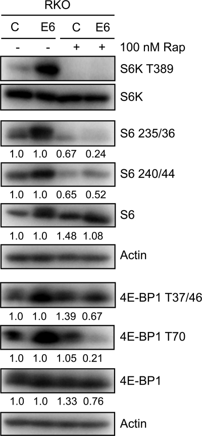
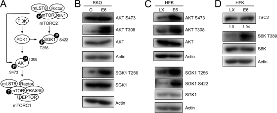
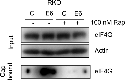
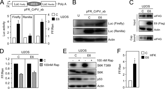
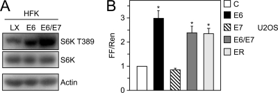
The mammalian target of rapamycin (mTOR) kinase acts as a cellular rheostat that integrates signals from a variety of cellular signal transduction pathways that sense growth factor and nutrient availability as well as intracellular energy status. It was previously reported that the human papillomavirus type 16 (HPV16) E6 oncoprotein may activate the S6 protein kinase (S6K) through binding and E6AP-mediated degradation of the mTOR inhibitor tuberous sclerosis complex 2 (TSC2) (Z. Lu, X. Hu, Y. Li, L. Zheng, Y. Zhou, H. Jiang, T. Ning, Z. Basang, C. Zhang, and Y. Ke, J. Biol. Chem. 279:35664-35670, 2004; L. Zheng, H. Ding, Z. Lu, Y. Li, Y. Pan, T. Ning, and Y. Ke, Genes Cells 13:285-294, 2008). Our results confirmed that HPV16 E6 expression causes an increase in mTORC1 activity through enhanced phosphorylation of mTOR and activation of downstream signaling pathways S6K and eukaryotic initiation factor binding protein 1 (4E-BP1). However, we did not detect a decrease in TSC2 levels in HPV16 E6-expressing cells. We discovered, however, that HPV16 E6 expression causes AKT activation through the upstream kinases PDK1 and mTORC2 under conditions of nutrient deprivation. We show that HPV16 E6 expression causes an increase in protein synthesis by enhancing translation initiation complex assembly at the 5' mRNA cap and an increase in cap-dependent translation. The increase in cap-dependent translation likely results from HPV16 E6-induced AKT/mTORC1 activation, as the assembly of the translation initiation complex and cap-dependent translation are rapamycin sensitive. Lastly, coexpression of the HPV16 E6 and E7 oncoproteins does not affect HPV16 E6-induced activation of mTORC1 and cap-dependent translation. HPV16 E6-mediated activation of mTORC1 signaling and cap-dependent translation may be a mechanism to promote viral replication under conditions of limited nutrient supply in differentiated, HPV oncoprotein-expressing proliferating cells.


Figures

FIG. 1.
FIG. 1.
HPV16 E6 expression activates mTOR1, 4E-BP1, S6K, and S6 phosphorylation through a TSC2-independent mechanism. (A) Schematic diagram of mTORC1 signaling. See text for details. (B to E) Western blot analysis of mTOR phosphorylation (B), TSC2 expression (with quantifications shown below) (C), 4E-BP1 phosphorylation (D), and S6K and S6 phosphorylation (E) in HPV16 E6-expressing and control RKO cells. A p53 blot is shown in panel B to document HPV16 E6 expression, and actin blots are shown as loading controls. Also shown are results from Western blot analysis of 4E-BP1 (F) and TSC2 expression and S6K and S6 phosphorylation (G) (with quantifications shown below) in HPV16 E6-expressing and control (LX) primary human foreskin keratinocyte cultures (HFKs). A p53 blot is shown in panel F to document HPV16 E6 expression, and actin blots are shown as loading controls.
FIG. 2.
FIG. 2.
HPV16 E6 expression causes increased S6K, S6, and 4E-BP1 phosphorylation through mTORC1 activation. Western blot analysis of mTORC1 downstream signaling components in RKO control and HPV16 E6-expressing RKO cells, treated with dimethyl sulfoxide (DMSO) or 100 nM rapamycin (Rap) for 1 h prior to lysis. Relative levels of unphosphorylated and phosphorylated species of S6 and 4E-BP1 are indicated. Actin blots are shown as loading controls.
FIG. 3.
FIG. 3.
HPV16 E6 expression causes AKT activation. (A) Schematic diagram of AKT phosphorylation through PDK1 and mTORC2 pathways. (B) Western blot analysis of AKT phosphorylation in control and HPV16 E6-expressing RKO cells. SGK1 is phosphorylated by PDK1 at T256 and is included as a control for PDK1 activation in HPV16 E6-expressing RKO cells. Actin blots are shown as loading controls. (C) Sustained AKT activation in control (LX) and HPV16 E6-expressing HFK populations under conditions of nutrient deprivation. SGK1 is phosphorylated by PDK1 at T256 and by mTORC2 at S422 and is included as a control for PDK1 and mTORC2 activation in HPV16 E6-expressing HFKs. Actin blots are shown as loading controls. (D) Sustained S6K activation in control (LX) and HPV16 E6-expressing HFK populations under conditions of nutrient deprivation. A TSC2 blot with quantification is shown to document similar expression in the two cell populations after nutrient deprivation; an actin blot is shown as a loading control.
FIG. 4.
FIG. 4.
Increased binding of the translation initiation factor eIF4G to a synthetic 7-methyl-GTP (7MeGTP) mRNA cap structure in HPV16 E6-expressing RKO cell lysates, which is sensitive to rapamycin treatment. Control and HPV16 E6-expressing RKO cells were treated with dimethyl sulfoxide (DMSO) or 100 nM rapamycin (Rap) for 1 h prior to lysis. Cap binding assays were performed as described in Materials and Methods. Levels of eIF4G in a 50-μg sample, representing 25% of the cap-binding reaction, together with an actin blot, are shown in the top panel (Input). Blot results for cap-bound eIF4G are shown in the bottom panel.
FIG. 5.
FIG. 5.
HPV16 E6 expression causes as increase in cap-dependent translation, which is sensitive to rapamycin treatment. (A) Diagram of bicistronic firefly Renilla reporter plasmid, pFR_CrPV_xb, used for these experiments. Firefly luciferase is translated through a cap-dependent mechanism, whereas Renilla luciferase is expressed from an internal ribosomal entry site (IRES) through a cap-independent mechanism (top). HPV16 E6 expression causes an increase in firefly but not Renilla luciferase activity (bottom). U2OS cells were transfected with control or HPV16 E6 expression vector, and lysates were processed for Renilla and firefly luciferase assays at 48 h posttransfection. The data are presented as the change of firefly and Renilla luciferase activities normalized to control vector-transfected cells (left and middle) and the fold change of normalized firefly compared to normalized Renilla luciferase activity (FF/Ren) (right). The bar graphs represent averages and standard deviations of four experiments, each performed in triplicate. The asterisk denotes statistical significance (P < 0.0001). (B) Western blot analysis of firefly and Renilla luciferase expression in U2OS cells transiently transfected with the indicated plasmids. U, untransfected cells. (C) Western blot analysis of eIF4G binding to a synthetic 7-methyl-GTP (7MeGTP) mRNA cap upon transient transfection of HPV16 E6 or control vector in U2OS cells. (D) HPV16 E6-mediated increase in cap-dependent translation is rapamycin sensitive. U2OS cells were transfected with pFR_CrPV_xb and the indicated plasmids; 18 h prior to lysis, cells were treated with dimethyl sulfoxide (DMSO) or 100 nM rapamycin (Rap). The graph represents averages and standard deviations of four experiments, each performed in triplicate. (E) Western blot analysis of S6K phosphorylation in U2OS cells transiently transfected with HPV16 E6 or control vector. One hour prior to lysis, cells were treated with DMSO or 100 nM rapamycin (Rap). Decreases in p53 levels are shown to document HPV16 E6 expression, and an actin blot is included as a loading control. (F) Transient transfection of HPV16 E6 activates cap-dependent translation in primary HFKs. Cells were transfected with pFR_CrPV_xb and the indicated plasmids and processed for Renilla and firefly luciferase assays at 48 h posttransfection. Firefly and Renilla luciferase activities were normalized to control vector-transfected cells and are presented as fold changes of normalized firefly relative to normalized Renilla luciferase activity. The bar graph represents the average and standard deviation of four experiments, each performed in triplicate; asterisks indicate statistical significance (P = 0.0001).
FIG. 6.
FIG. 6.
HPV16 E7 coexpression does not affect E6-induced S6K T389 phosphorylation or cap-dependent translation. (A) Western blot analysis of S6K T389 phosphorylation in HFK populations with stable expression of HPV16 E6 or HPV16 E6/E7 or control vector (LX)-transduced HFKs. An actin blot is shown as a loading control. (B) U2OS cells were transiently transfected with pFR_CrPV_xb and human β-actin-promoter-driven expression vectors for HPV16 E6, E7, E6/E7, the entire HPV16 early coding region (ER), or empty vector as a control and processed for Renilla and firefly luciferase assays at 48 h posttransfection. Firefly and Renilla luciferase activities were normalized to control vector-transfected cells and are presented as fold changes of normalized firefly relative to normalized Renilla luciferase activity. The bar represents the average and standard deviation of four experiments, each performed in triplicate; asterisks indicate statistical significance (P ≤ 0.0013).

References

    1. Alessi, D. R., S. R. James, C. P. Downes, A. B. Holmes, P. R. Gaffney, C. B. Reese, and P. Cohen. 1997. Characterization of a 3-phosphoinositide-dependent protein kinase which phosphorylates and activates protein kinase Bα. Curr. Biol. 7:261-269. - PubMed
    1. Baker, S. J., S. Markowitz, E. R. Fearon, J. K. Willson, and B. Vogelstein. 1990. Suppression of human colorectal carcinoma cell growth by wild-type p53. Science 249:912-915. - PubMed
    1. Biondi, R. M., A. Kieloch, R. A. Currie, M. Deak, and D. R. Alessi. 2001. The PIF-binding pocket in PDK1 is essential for activation of S6K and SGK, but not PKB. EMBO J. 20:4380-4390. - PMC - PubMed
    1. Budanov, A. V., and M. Karin. 2008. p53 target genes sestrin1 and sestrin2 connect genotoxic stress and mTOR signaling. Cell 134:451-460. - PMC - PubMed
    1. Charette, S. T., and D. J. McCance. 2007. The E7 protein from human papillomavirus type 16 enhances keratinocyte migration in an Akt-dependent manner. Oncogene 26:7386-7390. - PMC - PubMed

Publication types

MeSH terms

Substances