Review
. 2010 Oct 1;88(1):16-29.
doi: 10.1093/cvr/cvq237. Epub 2010 Jul 14.

Mitochondrial morphology and cardiovascular disease

Affiliations
Review

Mitochondrial morphology and cardiovascular disease

Sang-Bing Ong et al. Cardiovasc Res. .

Abstract

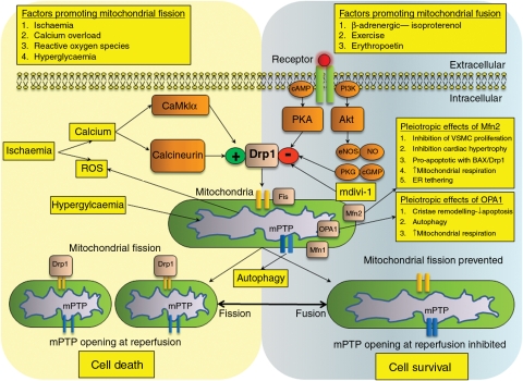
Mitochondria are dynamic and are able to interchange their morphology between elongated interconnected mitochondrial networks and a fragmented disconnected arrangement by the processes of mitochondrial fusion and fission, respectively. Changes in mitochondrial morphology are regulated by the mitochondrial fusion proteins (mitofusins 1 and 2, and optic atrophy 1) and the mitochondrial fission proteins (dynamin-related peptide 1 and mitochondrial fission protein 1) and have been implicated in a variety of biological processes including embryonic development, metabolism, apoptosis, and autophagy, although the majority of studies have been largely confined to non-cardiac cells. Despite the unique arrangement of mitochondria in the adult heart, emerging data suggest that changes in mitochondrial morphology may be relevant to various aspects of cardiovascular biology-these include cardiac development, the response to ischaemia-reperfusion injury, heart failure, diabetes mellitus, and apoptosis. Interestingly, the machinery required for altering mitochondrial shape in terms of the mitochondrial fusion and fission proteins are all present in the adult heart, but their physiological function remains unclear. In this article, we review the current developments in this exciting new field of mitochondrial biology, the implications for cardiovascular physiology, and the potential for discovering novel therapeutic strategies for treating cardiovascular disease.


Figures

Figure 1
Figure 1
Mitochondrial fission. The mitochondrial fission protein, Drp1, is located mainly in the cytosol and comprises a GTPase, a central region, and a GTPase effector domain (GED) or assembly domain. Fis1 is localized in the OMM with most of the protein facing into the cytosol, acting as a docking station for Drp1. On activation, Drp1 translocates to the mitochondria (a process which is regulated by phosphorylation and sumoylation), oligomerizes, and constricts the mitochondrial scission site, a process which requires GTPase, thereby resulting in mitochondrial division.
Figure 2
Figure 2
(A) Mitochondrial fusion. The mitochondrial fusion proteins Mfn1 and Mfn2 are located on the OMM with a cytosolic GTPase domain and two hydrophobic heptad repeat (HR) regions separated by a transmembrane repeat. The COOH-terminal HR region (HR2) mediates oligomerization between Mfn molecules on adjacent mitochondria allowing the membranes to fuse. GTP hydrolysis facilitates the fusion process. (B) OPA1 structure. The mitochondrial fusion protein, OPA1, comprises an N-terminal mitochondrial import sequence (MIS), hydrophobic heptad repeat (HR) segments, coiled-coil domain (C C), GTPase domain, a central domain, and a GTPase effector domain (GED) at the C terminus. OPA1 mediates the fusion of the inner mitochondrial membranes.
Figure 3
Figure 3
Representative confocal images of HL-1 cardiac cells transfected with mitochondrial red fluorescent protein depicting: (A) a cell displaying relatively fragmented mitochondria and (B) a cell displaying relatively elongated mitochondria.
Figure 4
Figure 4
Representative electron micrograph of adult murine heart depicting the three subpopulations of mitochondria: peri-nuclear (PN) mitochondria which are freely arranged in an area adjacent to the nucleus; interfibrillar (IF) mitochondria which are arranged along the myofibrils alongside the sarcomere; subsarcolemmal mitochondria (SSM) which are freely arranged in an area located just beneath the subsarcolemma.
Figure 5
Figure 5
(A and B) Representative electron micrographs of adult murine heart depicting elongated IF mitochondria marked with an asterisk, extending two to three sarcomeres in length. (C and D) Representative electron micrographs of adult murine heart following an episode of mild ischaemia (20 min), depicting fragmented rounded IF mitochondria. Of note, there is one elongated mitochondrion present on (D) marked with an asterisk.
Figure 6
Figure 6
Hypothetical scheme highlighting the potential roles of mitochondrial morphology in cardiovascular disease with factors promoting mitochondrial fission on the left of the figure and factors promoting mitochondrial fusion on the right of the figure. Central to the control of mitochondrial fission is the protein, Drp1, whose activity is regulated by phosphorylation. Cytosolic calcium overload (such as in ischaemia) induces mitochondrial fission by two mechanisms: (i) increasing the activity of calcineurin mediated dephosphorylation of Drp1 at Ser637 or (2) by increasing the activity of CaMkII which phosphorylates Drp1 at Ser637. In addition, in both the diabetic heart and the non-diabetic heart, hyperglycaemia induces mitochondrial fission through Drp1,, through a mechanism which is dependent on reactive oxygen species (ROS). Mitochondrial fission results in mitochondria which are more susceptible to mPTP opening, which results in cell death at the time of myocardial reperfusion. The phosphorylation of Drp1 at Ser637 by PKA prevents the translocation of Drp1 to the mitochondria and inhibits the process of mitochondrial fission. PKA can be activated using cAMP analogues, or by exercise or β-adrenergic stimulation using isoproterenol., Our recent preliminary data suggest that the cytokine erythropoietin can also induce mitochondrial fusion through the activation of Akt in cardiac cells, but the mechanism downstream of Akt remains underdetermined. However, one downstream target of Akt is the eNOS–NO–cGMP–PKG pathway which has recently been reported to phosphorylate and inhibit the pro-fission activity of Drp1. The mitochondrial division inhibitor-1 (mdivi-1) is a recently described small molecule inhibitor of Drp1, which has been demonstrated to limit both myocardial and renal ischaemic injury. In addition, the over-expression of Mfn1, Mfn2, and Drp1K38A can inhibit mitochondrial fission and promote survival in a cardiac cell line. However, both OPA1 and Mfn2 have a number of pleiotropic effects which appear independent of their pro-fusion effects. The inhibition of mitochondrial fission or the promotion of mitochondrial fusion prevents mPTP opening at reperfusion promoting cell survival. Recently, the mPTP has been demonstrated to play a role in signalling to the cell that a mitochondrion needs to be removed by autophagy.

References

    1. Hausenloy DJ, Scorrano L. Targeting cell death. Clin Pharmacol Ther. 2007;82:370–373. doi:10.1038/sj.clpt.6100352. - DOI - PubMed
    1. Liesa M, Palacin M, Zorzano A. Mitochondrial dynamics in mammalian health and disease. Physiol Rev. 2009;89:799–845. doi:10.1152/physrev.00030.2008. - DOI - PubMed
    1. Benda C. Ueber dier Spermatogenese de Verbebraten und hoherer Evertebraten, II. Theil: Die Histiogenese der Spermien. Arch Anat Physiol. 1898;73:393–398.
    1. Dimmer KS, Scorrano L. (De)constructing mitochondria: what for? Physiology (Bethesda) 2006;21:233–241. - PubMed
    1. Smirnova E, Shurland DL, Ryazantsev SN, van der Bliek AM. A human dynamin-related protein controls the distribution of mitochondria. J Cell Biol. 1998;143:351–358. doi:10.1083/jcb.143.2.351. - DOI - PMC - PubMed

Publication types

Substances