. 2010 Jul 14;30(28):9578-87.
doi: 10.1523/JNEUROSCI.2289-10.2010.

Efferent pathways modulate hyperactivity in inferior colliculus

Affiliations

Efferent pathways modulate hyperactivity in inferior colliculus

Wilhelmina Henrica A M Mulders et al. J Neurosci. .

Abstract

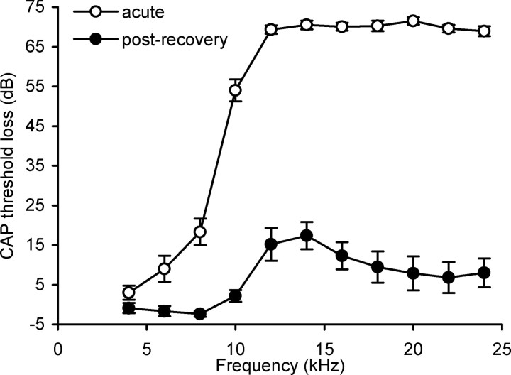
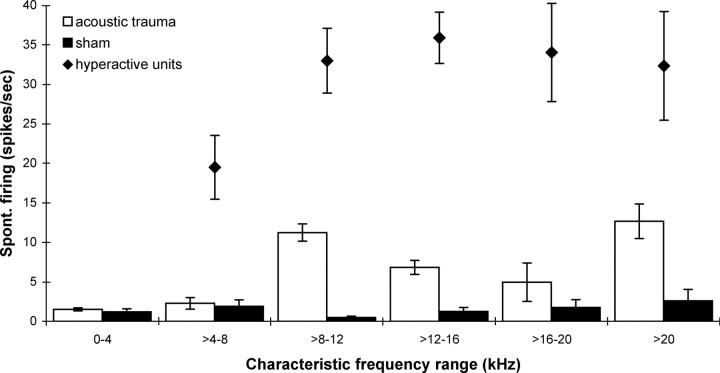
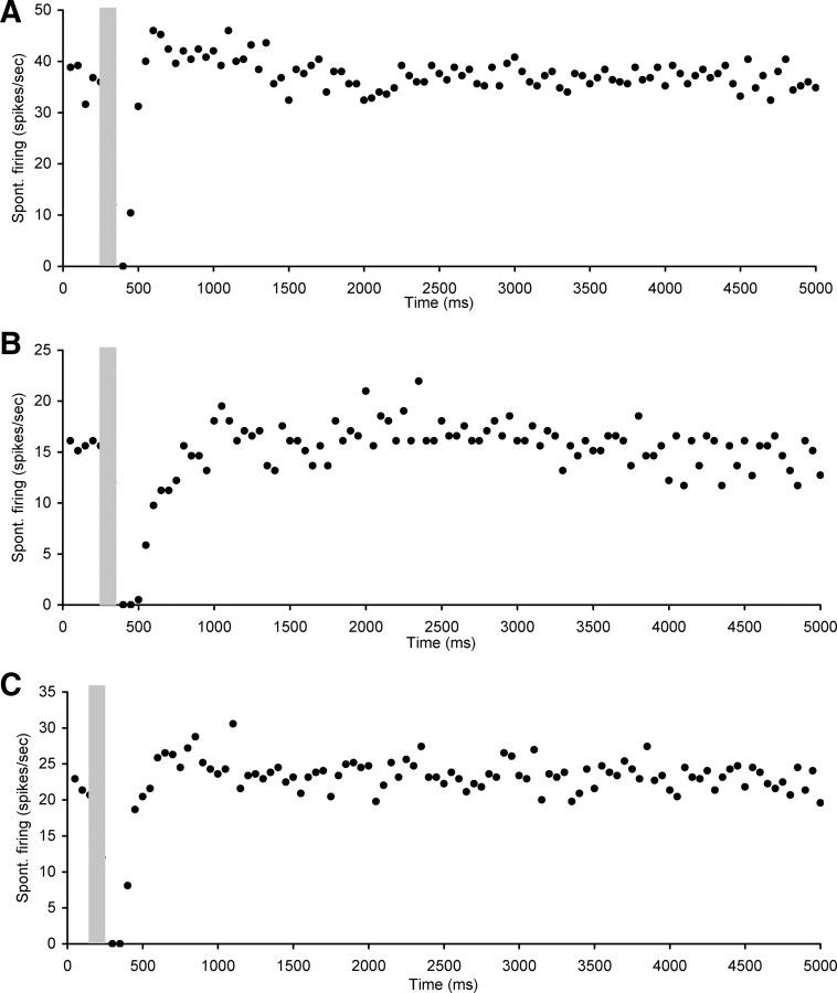
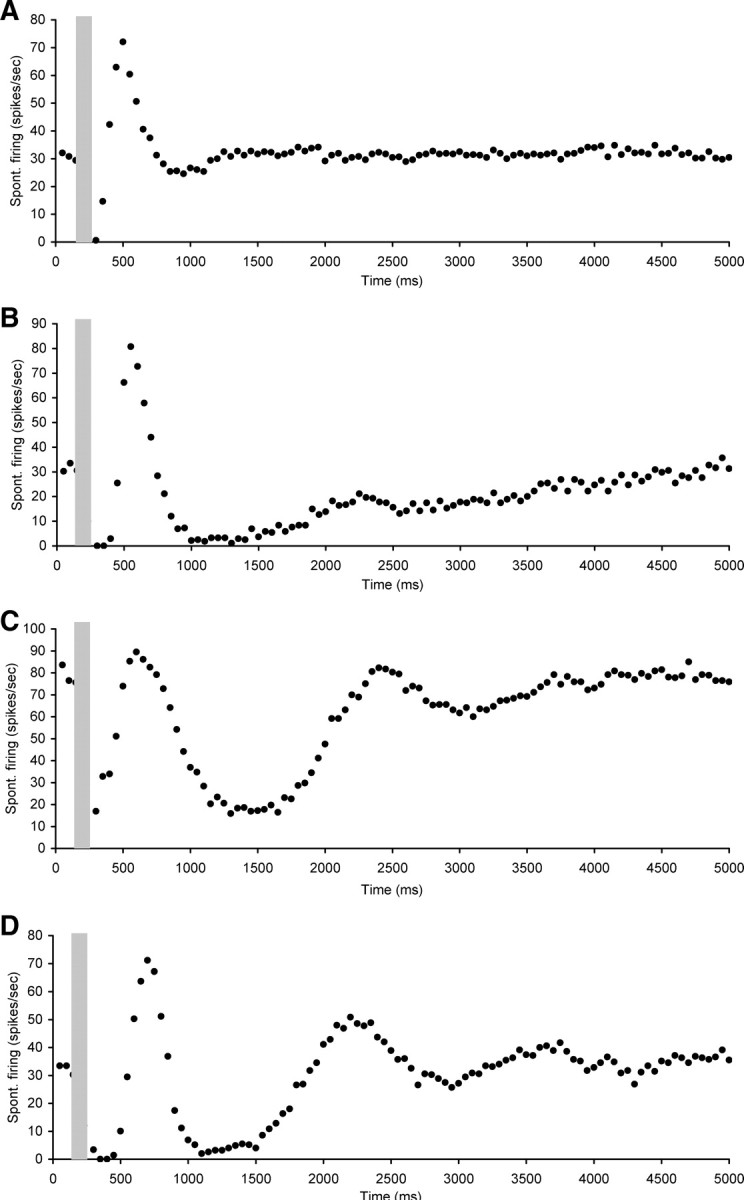
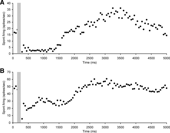
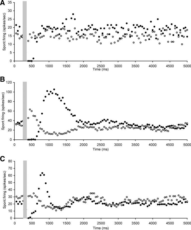
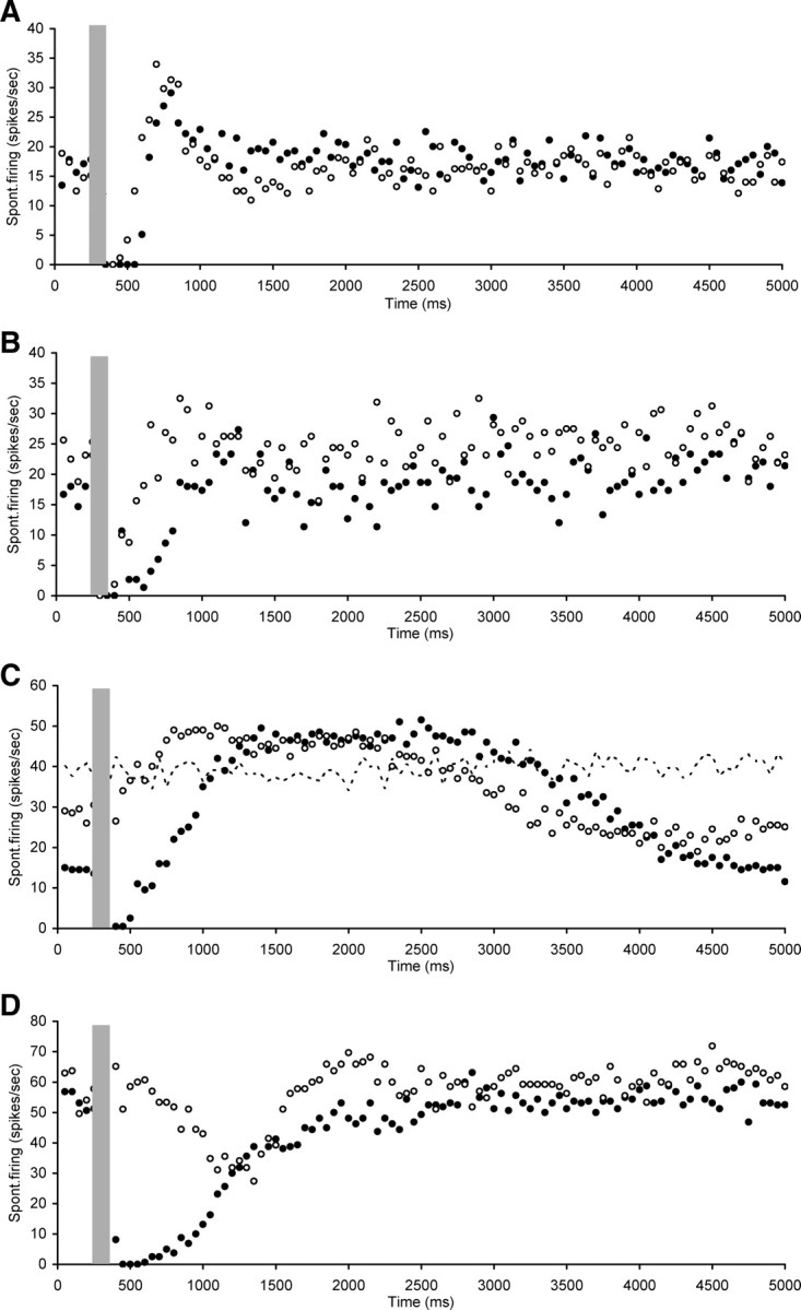
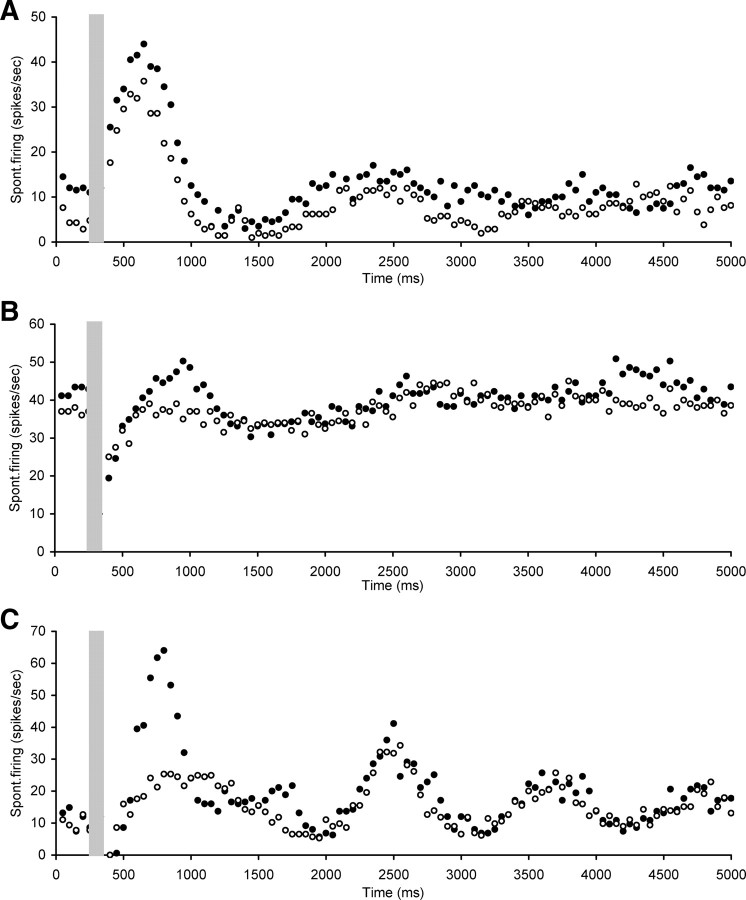
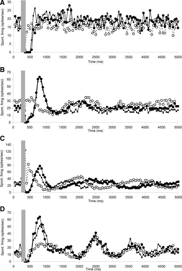
Animal models have demonstrated that mild hearing loss caused by acoustic trauma results in spontaneous hyperactivity in the central auditory pathways. This hyperactivity has been hypothesized to be involved in the generation of tinnitus, a phantom auditory sensation. We have recently shown that such hyperactivity, recorded in the inferior colliculus, is still dependent on cochlear neural output for some time after recovery (up to 6 weeks). We have now studied the capacity of an intrinsic efferent system, i.e., the olivocochlear system, to alter hyperactivity. This system is known to modulate cochlear neural output. Anesthetized guinea pigs were exposed to a loud sound and after 2 or 3 weeks of recovery, single-neuron recordings in inferior colliculus were made to confirm hyperactivity. Olivocochlear axons were electrically stimulated and effects on cochlear neural output and on highly spontaneous neurons in inferior colliculus were assessed. Olivocochlear stimulation suppressed spontaneous hyperactivity in the inferior colliculus. This result is in agreement with our earlier finding that hyperactivity can be modulated by altering cochlear neural output. Interestingly, the central suppression was generally much larger and longer lasting than reported previously for primary afferents. Blockade of the intracochlear effects of olivocochlear system activation eliminated some but not all of the effects observed on spontaneous activity, suggesting also a central component to the effects of stimulation. More research is needed to investigate whether these central effects of olivocochlear efferent stimulation are due to central intrinsic circuitry or to coactivation of central efferent collaterals to the cochlear nucleus.


Figures

Figure 1.
Figure 1.
Loss of cochlear sensitivity measured as CAP threshold change recorded in the cochlea after acute acoustic trauma (open circles; n = 16) and after recovery from acoustic trauma (black circles; n = 16). Excluded from this figure are three animals in which only the audiogram after recovery was measured. Data are mean ± SEM.
Figure 2.
Figure 2.
Averaged spontaneous firing rates per CF range in CNIC neurons in sham operated animals (black bars, n = 4) and in animals 2 weeks after acoustic trauma [open bars, n = 5, data taken from Mulders and Robertson (2009)]. Solid diamonds show averaged spontaneous firing rates of neurons selected for investigation of OCB effects. All data are mean ± SEM.
Figure 3.
Figure 3.
Scatter plots of spontaneous firing of hyperactive CNIC neurons with electrical stimulation of the OCB (gray bar), showing examples of short (ranging from 100 to 400 ms) suppression of spontaneous rate followed by a return to baseline spontaneous rate levels. A, Neuron CF 28 kHz, threshold 60 dB SPL. B, Neuron CF 6 kHz, threshold 22 dB SPL. C, Neuron CF 7.8 kHz, threshold 17 dB SPL. Stimulation parameters: 100 ms duration, 0.1 ms pulses, 300 Hz. Stimulation strength in A and B was 250 μA and in C 400 μA.
Figure 4.
Figure 4.
Scatter plots of spontaneous firing of hyperactive CNIC neurons with electrical stimulation of the OCB (gray bar), showing examples of short or nondetectable (ranging from 0 to 450 ms) suppression of spontaneous rate followed by more complex changes. A, Neuron CF 21.2 kHz, threshold 21 dB SPL. B, Neuron CF 10.9 kHz, threshold 29 dB SPL. C, Neuron CF 18.3 kHz, threshold 75 dB SPL. D, Neuron CF 9.7 kHz, threshold 28 dB SPL. Stimulation parameters: 100 ms duration, 0.1 ms pulses, 300 Hz. Stimulation strength in A was 350 μA, in B and C 250 μA, and in D 400 μA.
Figure 5.
Figure 5.
Scatter plots of spontaneous firing of hyperactive CNIC neurons with electrical stimulation of the OCB (gray bar), showing examples of long suppression after stimulation (ranging from 500 to 2550 ms) with either some late oscillation visible or a slow return to baseline levels. A, Neuron CF 18.6 kHz, threshold 82 dB SPL. B, Neuron CF 10.9 kHz, threshold 56 dB SPL. Stimulation parameters: 100 ms duration, 0.1 ms pulses, 300 Hz. Stimulation strength in A and B was 250 μA.
Figure 6.
Figure 6.
Scatter plots of spontaneous firing of hyperactive CNIC neurons with electrical stimulation of the OCB (gray bar), recorded both before (dark circles) and after (open circles) strychnine perfusion in the cochlea. Illustrations of neurons where the immediate suppression was abolished (A–C), but sometimes later effects remained (B, C). A, Neuron CF 7.8 kHz, threshold 20 dB SPL. B, Neuron CF 9.2 kHz, threshold 21 dB SPL. C, Neuron CF 14.5 kHz, threshold 22 dB SPL. Stimulation parameters: 100 ms duration, 0.1 ms pulses, 300 Hz. Stimulation strength in A and B was 300 μA and in C 150 μA.
Figure 7.
Figure 7.
Scatter plots of spontaneous firing of hyperactive CNIC neurons with electrical stimulation of the OCB (gray bar), recorded both before (dark circles) and after (open circles) strychnine perfusion in the cochlea. Illustrations of neurons in which the immediate suppression observed after OCB stimulation was not completely abolished by cochlear strychnine perfusion but its duration was reduced. In C also indicated is the spontaneous firing rate without OCB stimulation or strychnine perfusion (dotted line). A, Neuron CF 8.5 kHz, threshold 43 dB SPL. B, Neuron CF 9.2 kHz, threshold 67 dB SPL. C, Neuron nonresponsive to sound. D, Neuron CF 9 kHz, threshold 61 dB SPL. Stimulation parameters: 100 ms duration, 0.1 ms pulses, 300 Hz. Stimulation strength in A and D was 250 μA and in B and C 300 μA.
Figure 8.
Figure 8.
Scatter plots of spontaneous firing of hyperactive CNIC neurons with electrical stimulation of the OCB (gray bar), recorded both before (dark circles) and after (open circles) strychnine perfusion in the cochlea. A, Illustration of neuron in which strychnine perfusion did not change effects of OCB stimulation. B, C, Two neurons in which strychnine perfusion in the cochlea did not affect the suppression, but did decrease the following overshoot. A, Neuron CF 22.9 kHz, threshold 44 dB SPL. B, Neuron CF 18 kHz, threshold 66 dB SPL. C, Neuron CF 16.8 kHz, threshold 36 dB SPL. Stimulation parameters: 100 ms duration, 0.1 ms pulses, 300 Hz. Stimulation strength in A was 250 μA, in B 350 μA, and in C 150 μA.
Figure 9.
Figure 9.
Scatter plots of spontaneous firing of hyperactive CNIC neurons with electrical stimulation of the OCB (gray bar), recorded before (dark circles) and after (open circles) strychnine perfusion in the cochlea, as well as after recovery from the strychnine effects (dark triangles). Note reversibility of the strychnine effects and the repeatability of all components of the OCB stimulation effects before blockade and after recovery. A, Neuron CF 7.8 kHz, threshold 20 dB SPL. B, Neuron CF 14.5 kHz, threshold 23 dB SPL. C, Neuron CF 13.9 kHz, threshold 39 dB SPL. D, Neuron CF 16.8 kHz, threshold 36 dB SPL. Stimulation parameters: 100 ms duration, 0.1 ms pulses, 300 Hz. Stimulation strength in A was 250 μA and in B–D 150 μA.

References

    1. Arnott RH, Wallace MN, Shackleton TM, Palmer AR. Onset neurones in the anteroventral cochlear nucleus project to the dorsal cochlear nucleus. J Assoc Res Otolaryngol. 2004;5:153–170. - PMC - PubMed
    1. Bauer CA, Turner JG, Caspary DM, Myers KS, Brozoski TJ. Tinnitus and inferior colliculus activity in chinchillas related to three distinct patterns of cochlear trauma. J Neurosci Res. 2008;86:2564–2578. - PMC - PubMed
    1. Benson CG, Potashner SJ. Retrograde transport of [3H]glycine from the cochlear nucleus to the superior olive in the guinea pig. J Comp Neurol. 1990;296:415–426. - PubMed
    1. Benson TE, Brown MC. Synapses formed by olivocochlear axon branches in the mouse cochlear nucleus. J Comp Neurol. 1990;295:52–70. - PubMed
    1. Benson TE, Berglund AM, Brown MC. Synaptic input to cochlear nucleus dendrites that receive medial olivocochlear synapses. J Comp Neurol. 1996;365:27–41. - PubMed

Publication types

LinkOut - more resources