. 2010 Jun 24:4:16.
doi: 10.3389/fnbeh.2010.00016. eCollection 2010.

How neurons generate behavior in a hatchling amphibian tadpole: an outline

Affiliations

How neurons generate behavior in a hatchling amphibian tadpole: an outline

Alan Roberts et al. Front Behav Neurosci. .

Abstract

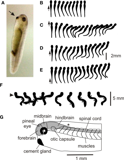
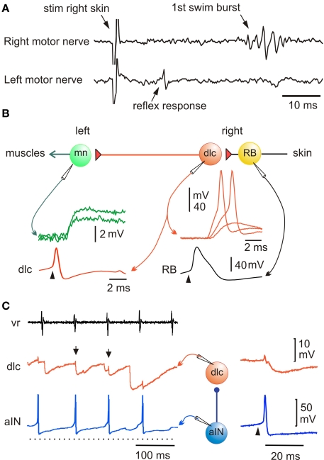
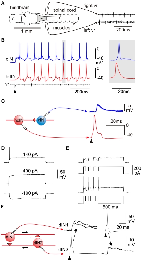
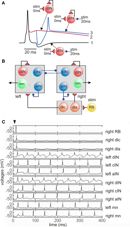
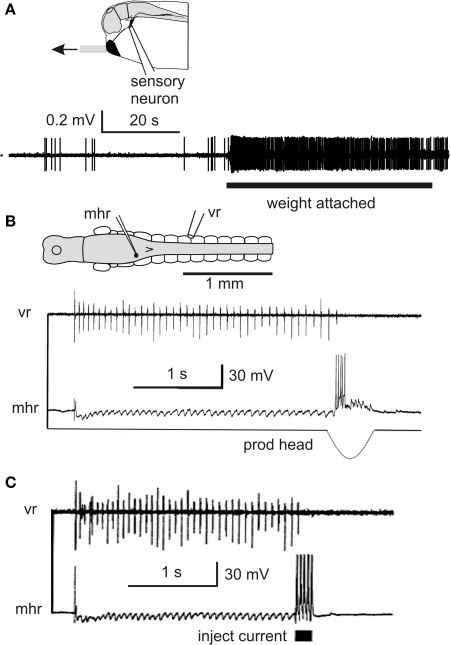
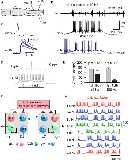
Adult nervous systems are so complex that understanding how they produce behavior remains a real challenge. We chose to study hatchling Xenopus tadpoles where behavior is controlled by a few thousand neurons but there is a very limited number of types of neuron. Young tadpoles can flex, swim away, adjust their trajectory, speed-up and slow-down, stop when they contact support and struggle when grasped. They are sensitive to touch, pressure, noxious stimuli, light intensity and water currents. Using whole-cell recording has led to rapid progress in understanding central networks controlling behavior. Our methods are illustrated by an analysis of the flexion reflex to skin touch. We then define the seven types of neuron that allow the tadpole to swim when the skin is touched and use paired recordings to investigate neuron properties, synaptic connections and activity patterns. Proposals on how the swim network operates are evaluated by experiment and network modeling. We then examine GABAergic inhibitory pathways that control swimming but also produce tonic inhibition to reduce responsiveness when the tadpole is at rest. Finally, we analyze the strong alternating struggling movements the tadpole makes when grasped. We show that the mechanisms for rhythm generation here are very different to those during swimming. Although much remains to be explained, study of this simple vertebrate has uncovered basic principles about the function and organization of vertebrate nervous systems.

Keywords: Xenopus; pattern generation; reflex; spinal interneurons; tonic inhibition.


Figures

Figure 1
Figure 1
Hatchling Xenopus tadpole and its behavior. (A) Stage 37/38 tadpole (∼5 mm long) at rest hanging by mucus from its cement gland (arrow) attached to the glass side of a tank. (B–E) Response to a short stroke with a hair (small arrow) seen from above in movies at 200 fps. (based on Boothby and Roberts, 1995) (B) Flexion (C–E) swimming (F) struggling when held at the neck (arrow). Points of maximum bending are shown by dots and circles, 64 fps. (based on Kahn and Roberts, 1982b). (G) Side view of the head end to show the main CNS regions.
Figure 2
Figure 2
Pathway for a simple flexion reflex. (A) motor nerve recordings show that a skin stimulus on the right produces a short latency reflex response on the left. (B) Diagram of reflex pathway where sensory RB neurons excite dlc interneurons and they excite motoneurons on the opposite side. Paired recordings show that current to the RB (arrowhead) leads to an action potential and after delay, to a large EPSP or action potential in the dlc interneuron; an action potential in the dlc leads to a small EPSP in the motoneuron. (based on Li et al., 2003b). (C) A paired recording shows an aIN active during swimming (monitored by ventral root electrode, vr) and that some aIN spikes lead to IPSPs in a dlc interneuron (small arrows). If current is injected to make the aIN fire (right panel; arrowhead) an IPSP is seen in the dlc. (based on Li et al., 2002).
Figure 3
Figure 3
Swimming: effects of lesions, activity patterns and excitation. (A) CNS in dorsal view to show suction electrode positions to record alternating ventral root (vr) spikes during swimming generated by an isolated 0.3-mm long region of CNS (gray). (B) Whole-cell recording from a cIN and hindbrain dIN (hdIN) show both fire once on each cycle of swimming and the hdIN has a long action potential; (C) spike evoked in the hdIN by current (arrowhead) leads to an EPSP in the cIN. (D) A dIN fires once to positive current but does not fire on rebound after negative current. (E) If large enough, negative current pulses during depolarization can lead to post-inhibitory rebound firing. (F) Recording from a pair of dINs in the hindbrain shows that when current is injected to make them fire (arrowheads) they produce long duration excitation in the other. (based on Li et al., 2006).
Figure 4
Figure 4
Feedback excitation and post-inhibitory rebound hypothesis for the swimming circuit. (A) Responses of a single dIN illustrate the fundamental mechanisms. (1) Fast short excitation at time 0 makes dIN fire but short inhibition at 20 ms does not lead to rebound from the resting potential (black line). (2) when dIN feeds back long NMDA excitation, it holds the dIN depolarized but it does not fire (red). (3) if inhibition occurs during depolarization it leads to rebound dIN firing and a new cycle can start (blue). (B) The model network with populations equivalent to 30 of each type of neuron in each half-center. A skin stimulus to one RB neuron activates the network via sensory pathway interneurons. (C) Example of network swimming activity following a single RB impulse. (based on Sautois et al., 2007).
Figure 5
Figure 5
Inhibitory pathways stop swimming and reduce responses. (A) Impulse recording from trigeminal sensory neuron innervating the cement gland shows the resting discharge and increased firing when the cement gland mucus strand is in tension. (based on Lambert et al., 2004a) (B) Intracellular recording from an mhr, with long descending contralateral axon in spinal cord, shows it is inhibited during swimming and fires when the cement gland is prodded and swimming stops (C). Injecting current to make this single mhr fire can stop swimming. (based on Perrins et al., 2002).
Figure 6
Figure 6
The struggling motor pattern, the neurons responsible and its mechanism of generation. (A,B) During 40-Hz skin stimuli struggling is evoked and a cIN and ecIN recorded in whole-cell mode are recruited. When stimulation stops the motor pattern switches to swimming and recorded neurons become silent. (C) Current injection into the ecIN makes it fire an action potential which produces a short latency EPSP in the cIN. (D) Longer injected current induces typical repetitive firing. (E) Measures of 1st and 10th compound IPSCs evoked in a dIN by stimulation of the opposite side show significant depression only with 100-Hz stimulation. (F) Half-center model of struggling network without length with three of each type of neuron on each side. (G) If cIN inhibition shows depression, this network can reliably generate struggling- like activity during tonic sensory excitation. (based on Li et al., 2007).

References

    1. Aiken S. P., Kuenzi F. M., Dale N. (2003). Xenopus embryonic spinal neurons recorded in situ with patch-clamp electrodes–conditional oscillators after all?. Eur J. Neurosci. 18, 333–34310.1046/j.1460-9568.2003.02755.x - DOI - PubMed
    1. Arshavsky Y., Orlovsky G. N., Panchin Y. V., Roberts A., Soffe S. R. (1993). Neuronal control of swimming locomotion: analysis of the pteropod mollusc Clione and embryos of the amphibian Xenopus. Trends Neurosci. 16, 227–23310.1016/0166-2236(93)90161-E - DOI - PubMed
    1. Berg R. W., Alaburda A., Hounsgaard J. (2007). Balanced inhibition and excitation drive spike activity in spinal half-centers. Science 315, 390–39310.1126/science.1134960 - DOI - PubMed
    1. Berkowitz A., Roberts A., Soffe S. R. (2010). Roles for multifunctional and specialized spinal interneurons during motor pattern generation in tadpoles, zebrafish larvae, and turtles. Front. Behav. Neurosci 4:3610.3389/fnbeh.2010.00036 - DOI - PMC - PubMed
    1. Boothby K. M., Roberts A. (1992). The stopping response of Xenopus laevis embryos: pharmacology and intracellular physiology of rhythmic spinal neurones and hindbrain neurones. J. Exp. Biol. 169, 65–8610.1007/BF00196899 - DOI - PubMed

LinkOut - more resources