

doi: 10.1155/2010/456841.
Epub 2010 Jun 15.
Apoptotic pathways in pemphigus
Affiliations
- PMID: 20631907
- PMCID: PMC2902125
- DOI: 10.1155/2010/456841
Item in Clipboard
Apoptotic pathways in pemphigus
Dermatol Res Pract.
2010.
Abstract
Pemphigus is a group of human autoimmune blistering diseases of the skin in which autoantibodies to desmosome cadherins induce loss of cell-cell adhesion (acantholysis). In addition to steric hindrance and activation of intracellular signaling, apoptosis has been suggested to contribute to the mechanism by which pathogenic IgG induces acantholysis. We review the current literature examining the role of apoptosis in pemphigus. Current data suggest that apoptosis is not required for blister induction, but that activation of proapoptotic proteins, including caspase cysteine proteinases, may sensitize cells to the acantholytic effects of pemphigus IgG.
Figures
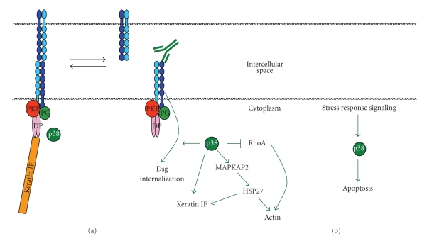
Pemphigus, p38MAPK, acantholysis and apoptosis. Two sequential peaks of p38MAPK activation are observed when keratinocytes are exposed to either PV or PF IgG. (a) Pemphigus IgG binds to dsg and biases the equilibrium of desmosome assembly/disassembly towards disassembly which is linked by an, as yet, undefined mechanism towards activation of p38MAPK. Subsequent p38 dependent alterations in the cell state include RhoA inactivation, dsg endocytosis, HSP27 phosphorylation, keratin intermediate filament retraction, actin, and loss of cell-cell adhesion (acantholysis). (b) A second late peak of p38 activity is observed that is likely a stress response signal induced by loss of cell-cell adhesion and leads to activation of proapoptotic pathways including caspase-3 activation.
Similar articles
-
Caspase cascade pathways of apoptosis in oral pemphigus: An immunohistochemical study.J Oral Maxillofac Pathol. 2018 Jan-Apr;22(1):48-53. doi: 10.4103/jomfp.JOMFP_79_17. J Oral Maxillofac Pathol. 2018. PMID: 29731556 Free PMC article.
-
Apoptotic pathways in the pathogenesis of pemphigus: targets for new therapies.Curr Pharm Biotechnol. 2012 Aug;13(10):1877-81. doi: 10.2174/138920112802273236. Curr Pharm Biotechnol. 2012. PMID: 22250711 Review.
-
Apoptotic mechanism in pemphigus autoimmunoglobulins-induced acantholysis--possible involvement of the EGF receptor.Autoimmunity. 2006 Nov;39(7):563-75. doi: 10.1080/08916930600971836. Autoimmunity. 2006. PMID: 17101500
-
Apoptosis in pemphigus.Autoimmun Rev. 2009 Jun;8(7):533-7. doi: 10.1016/j.autrev.2009.01.011. Epub 2009 Feb 2. Autoimmun Rev. 2009. PMID: 19189866 Review.
-
Apoptolysis: a novel mechanism of skin blistering in pemphigus vulgaris linking the apoptotic pathways to basal cell shrinkage and suprabasal acantholysis.Exp Dermatol. 2009 Sep;18(9):764-70. doi: 10.1111/j.1600-0625.2009.00934.x. Epub 2009 Jun 25. Exp Dermatol. 2009. PMID: 19555352
Cited by
-
The Evolving Story of Autoantibodies in Pemphigus Vulgaris: Development of the "Super Compensation Hypothesis".Front Med (Lausanne). 2018 Aug 14;5:218. doi: 10.3389/fmed.2018.00218. eCollection 2018. Front Med (Lausanne). 2018. PMID: 30155465 Free PMC article. Review.
-
Apoptosis in immune-mediated diseases.J Pharm Bioallied Sci. 2015 Apr;7(Suppl 1):S200-2. doi: 10.4103/0975-7406.155902. J Pharm Bioallied Sci. 2015. PMID: 26015710 Free PMC article. Review.
-
Autoantibody-Specific Signalling in Pemphigus.Front Med (Lausanne). 2021 Aug 9;8:701809. doi: 10.3389/fmed.2021.701809. eCollection 2021. Front Med (Lausanne). 2021. PMID: 34434944 Free PMC article. Review.
-
The caspase pathway as a possible therapeutic target in experimental pemphigus.Autoimmune Dis. 2011 Mar 2;2011:563091. doi: 10.4061/2011/563091. Autoimmune Dis. 2011. PMID: 21403857 Free PMC article.
-
Caspase cascade pathways of apoptosis in oral pemphigus: An immunohistochemical study.J Oral Maxillofac Pathol. 2018 Jan-Apr;22(1):48-53. doi: 10.4103/jomfp.JOMFP_79_17. J Oral Maxillofac Pathol. 2018. PMID: 29731556 Free PMC article.
References
-
- Amagai M, Klaus-Kovtun V, Stanley JR. Autoantibodies against a novel epithelial cadherin in Pemphigus vulgaris, a disease of cell adhesion. Cell. 1991;67(5):869–877. - PubMed
-
- Ding X, Aoki V, Mascaro JM, Lopez-Swiderski A, Diaz LA, Fairley JA. Mucosal and mucocutaneous (generalized) pemphigus vulgaris show distinct autoantibody profiles. Journal of Investigative Dermatology. 1997;109(4):592–596. - PubMed
-
- Amagai M, Tsunoda K, Zillikens D, Nagai T, Nishikawa T. The clinical phenotype of pemphigus is defined by the anti-desmoglein autoantibody profile. Journal of the American Academy of Dermatology. 1999;40(2, part 1):167–170. - PubMed
-
- Miyagawa S, Amagai M, Iida T, Yamamoto Y, Nishikawa T, Shirai T. Late development of antidesmoglein 1 antibodies in pemphigus vulgaris: correlation with disease progression. British Journal of Dermatology. 1999;141(6):1084–1087. - PubMed
Grants and funding
LinkOut - more resources
Full Text Sources
