

Fast 3D chemical exchange saturation transfer (CEST) imaging of the human brain
- PMID: 20632402
- PMCID: PMC2932772
- DOI: 10.1002/mrm.22546
Fast 3D chemical exchange saturation transfer (CEST) imaging of the human brain
Abstract
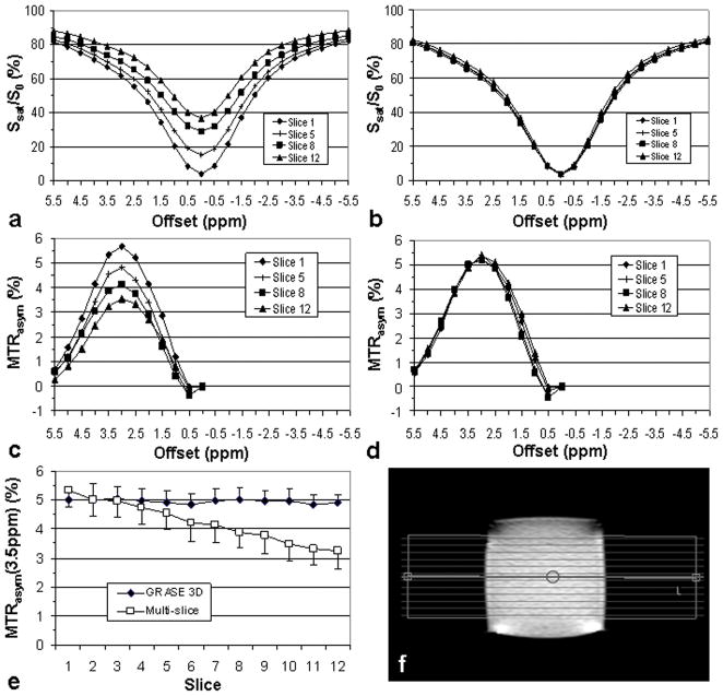
Chemical exchange saturation transfer magnetic resonance imaging can detect low-concentration compounds with exchangeable protons through saturation transfer to water. This technique is generally slow, as it requires acquisition of saturation images at multiple frequencies. In addition, multislice imaging is complicated by saturation effects differing from slice to slice because of relaxation losses. In this study, a fast three-dimensional chemical exchange saturation transfer imaging sequence is presented that allows whole-brain coverage for a frequency-dependent saturation spectrum (z-spectrum, 26 frequencies) in less than 10 min. The approach employs a three-dimensional gradient- and spin-echo readout using a prototype 32-channel phased-array coil, combined with two-dimensional sensitivity encoding accelerations. Results from a homogenous protein-containing phantom at 3T show that the sequence produced a uniform contrast across all slices. To show translational feasibility, scans were also performed on five healthy human subjects. Results for chemical exchange saturation transfer images at 3.5 ppm downfield of the water resonance, so-called amide proton transfer images, show that lipid signals are sufficiently suppressed and artifacts caused by B(0) inhomogeneity can be removed in postprocessing. The scan time and image quality of these in vivo results show that three-dimensional chemical exchange saturation transfer MRI using gradient- and spin-echo acquisition is feasible for whole-brain chemical exchange saturation transfer studies at 3T in a clinical time frame.
2010 Wiley-Liss, Inc.
Figures


Similar articles
-
Fast 3D chemical exchange saturation transfer imaging with variably-accelerated sensitivity encoding (vSENSE).Magn Reson Med. 2019 Dec;82(6):2046-2061. doi: 10.1002/mrm.27881. Epub 2019 Jul 1. Magn Reson Med. 2019. PMID: 31264278
-
In vivo three-dimensional whole-brain pulsed steady-state chemical exchange saturation transfer at 7 T.Magn Reson Med. 2012 Jun;67(6):1579-89. doi: 10.1002/mrm.23141. Epub 2011 Nov 14. Magn Reson Med. 2012. PMID: 22083645 Free PMC article.
-
3D gradient echo snapshot CEST MRI with low power saturation for human studies at 3T.Magn Reson Med. 2019 Apr;81(4):2412-2423. doi: 10.1002/mrm.27569. Epub 2018 Nov 15. Magn Reson Med. 2019. PMID: 30431179 Free PMC article.
-
Ytterbium chelated to 1,4,7,10-tetraazacyclododecane-1,4,7-triacetic acid,10-orthoaminoanilide.2011 Nov 26 [updated 2012 Jan 5]. In: Molecular Imaging and Contrast Agent Database (MICAD) [Internet]. Bethesda (MD): National Center for Biotechnology Information (US); 2004–2013. 2011 Nov 26 [updated 2012 Jan 5]. In: Molecular Imaging and Contrast Agent Database (MICAD) [Internet]. Bethesda (MD): National Center for Biotechnology Information (US); 2004–2013. PMID: 22238803 Free Books & Documents. Review.
-
Application of chemical exchange saturation transfer (CEST) MRI for endogenous contrast at 7 Tesla.J Neuroimaging. 2013 Oct;23(4):526-32. doi: 10.1111/j.1552-6569.2012.00751.x. Epub 2013 Feb 12. J Neuroimaging. 2013. PMID: 23402307 Free PMC article. Review.
Cited by
-
Development and Validation of Four Different Methods to Improve MRI-CEST Tumor pH Mapping in Presence of Fat.J Imaging. 2024 Jul 12;10(7):166. doi: 10.3390/jimaging10070166. J Imaging. 2024. PMID: 39057737 Free PMC article.
-
Radiomics analysis of amide proton transfer-weighted and structural MR images for treatment response assessment in malignant gliomas.NMR Biomed. 2023 Jan;36(1):e4824. doi: 10.1002/nbm.4824. Epub 2022 Sep 24. NMR Biomed. 2023. PMID: 36057449 Free PMC article.
-
Natural D-glucose as a biodegradable MRI contrast agent for detecting cancer.Magn Reson Med. 2012 Dec;68(6):1764-73. doi: 10.1002/mrm.24520. Epub 2012 Oct 16. Magn Reson Med. 2012. PMID: 23074027 Free PMC article.
-
Evaluating the Role of Amide Proton Transfer (APT)-Weighted Contrast, Optimized for Normalization and Region of Interest Selection, in Differentiation of Neoplastic and Infective Mass Lesions on 3T MRI.Mol Imaging Biol. 2020 Apr;22(2):384-396. doi: 10.1007/s11307-019-01382-x. Mol Imaging Biol. 2020. PMID: 31228076 Free PMC article.
-
Chemical exchange rotation transfer (CERT) on human brain at 3 Tesla.Magn Reson Med. 2018 Dec;80(6):2609-2617. doi: 10.1002/mrm.27365. Epub 2018 May 25. Magn Reson Med. 2018. PMID: 29802641 Free PMC article.
References
-
- Ward KM, Aletras AH, Balaban RS. A new class of contrast agents for MRI based on proton chemical exchange dependent saturation transfer (CEST) J Magn Reson. 2000;143:79–87. - PubMed
-
- Zhou J, van Zijl PC. Chemical exchange saturation transfer imaging and spectroscopy. Progr NMR Spectr. 2006;48:109–136.
-
- Aime S, Crich SG, Gianolio E, Giovenzana GB, Tei L, Terreno E. High sensitivity lanthanide(III) based probes for MR-medical imaging. Coord Chem Rev. 2006;250:1562–1579.
-
- Goffeney N, Bulte JWM, Duyn J, Bryant LH, van Zijl PCM. Sensitive NMR detection of cationic-polymer-based gene delivery systems using saturation transfer via proton exchange. J Am Chem Soc. 2001;123:8628–8629. - PubMed
Publication types
MeSH terms
Substances
Grants and funding
LinkOut - more resources
Full Text Sources
Other Literature Sources
