

Changes in cardiac pumping efficiency and intra-thoracic organ volume during negative pressure wound therapy of sternotomy wounds, assessment using magnetic resonance imaging
- PMID: 20633058
- PMCID: PMC7951601
- DOI: 10.1111/j.1742-481X.2010.00712.x
Changes in cardiac pumping efficiency and intra-thoracic organ volume during negative pressure wound therapy of sternotomy wounds, assessment using magnetic resonance imaging
Abstract
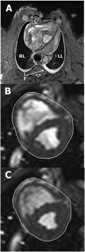
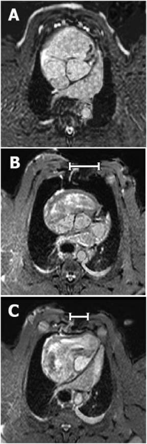
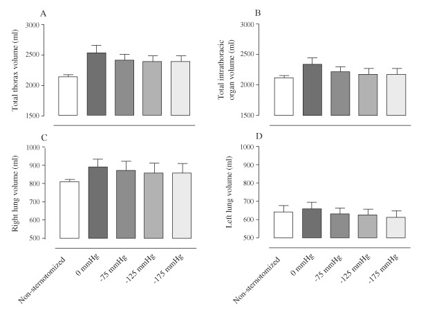
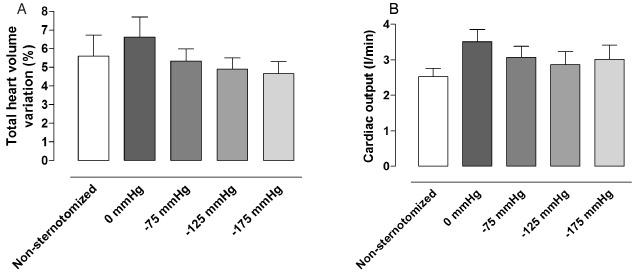
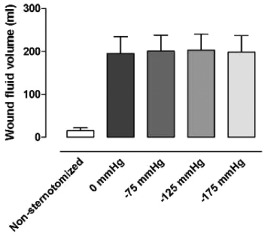
Knowledge on the effects of negative pressure wound therapy (NPWT) on the intra-thoracic organs is limited. The present study was performed to investigate the effects of NPWT on the volume of the intra-thoracic organs, using magnetic resonance imaging (MRI), in a porcine sternotomy wound model. Six pigs underwent median sternotomy followed by NPWT at -75, -125 and -175 mmHg. Six pigs were not sternotomised. MR images covering the thorax and heart were acquired. The volumes of the thoracic cavity, lungs, wound fluid and heart were then determined. The volumes of the thoracic cavity and intra-thoracic organs increased after sternotomy and decreased upon NPWT application. The total heart volume variation, which is inversely related to cardiac pumping efficiency, was higher after sternotomy and decreased during NPWT. NPWT did not result in the evacuation of wound fluid from the bottom of the wound. NPWT largely closes and restores the thoracic cavity. Cardiac pumping efficiency returns to pre-sternotomy levels during NPWT. This may contribute to the clinical benefits of NPWT over open-chest care, including the stabilizing effects and the reduced need for mechanical ventilation.
Figures
Erratum for
-
Changes in cardiac pumping efficiency and intra-thoracic organ volume during negative pressure wound therapy of sternotomy wounds, assessment using magnetic resonance imaging.Int Wound J. 2010 Apr;7(2):115-21. doi: 10.1111/j.1742-481X.2010.00664.x. Int Wound J. 2010. PMID: 20529152 Free PMC article.
References
-
- Torbrand C, Ugander M, Engblom H, Olivecrona GK, Gålne O, Arheden H, Ingemansson R, Malmsjö M. Changes in cardiac pumping efficiency and intra‐thoracic organ volume during negative pressure wound therapy of sternotomy wounds, assessment using magnetic resonance imaging. Int Wound J 2010;7:115–21. - PMC - PubMed
-
- Sjogren J, Gustafsson R, Nilsson J, Malmsjo M, Ingemansson R. Clinical outcome after poststernotomy mediastinitis: vacuum‐assisted closure versus conventional treatment. Ann Thorac Surg 2005;79:2049–55. - PubMed
-
- Malmsjo M, Ingemansson R, Sjogren J. Mechanisms governing the effects of vacuum‐assisted closure in cardiac surgery. Plast Reconstr Surg 2007;120:1266–75. - PubMed
-
- Ramnarine IR, McLean A, Pollock JC. Vacuum‐assisted closure in the paediatric patient with post‐cardiotomy mediastinitis. Eur J Cardiothorac Surg 2002;22:1029–31. - PubMed
-
- Kutschka I, Frauendorfer P, Harringer W. Vacuum assisted closure therapy improves early postoperative lung function in patients with large sternal wounds. Zentralbl Chir 2004;129 Suppl 1:S33–4. - PubMed
Publication types
MeSH terms
LinkOut - more resources
Full Text Sources
Medical
