. 2010 Jul 15:10:209.
doi: 10.1186/1471-2334-10-209.

Resistance of Leishmania (Viannia) braziliensis to nitric oxide: correlation with antimony therapy and TNF-alpha production

Affiliations

Resistance of Leishmania (Viannia) braziliensis to nitric oxide: correlation with antimony therapy and TNF-alpha production

Anselmo S Souza et al. BMC Infect Dis. .

Abstract

Background: Nitric oxide (NO) produced in macrophages plays a pivotal role as a leishmanicidal agent. A previous study has demonstrated that 20% of the L. (V.) braziliensis isolated from initial cutaneous lesions of patients from the endemic area of Corte de Pedra, Bahia, Brazil, were NO resistant. Additionally, 5 to 11% of the patients did not respond to three or more antimony treatments" (refractory patients). The aim of this study is to investigate if there is an association between the resistance of L. (V.) braziliensis to NO and nonresponsiveness to antimony therapy and cytokine production.

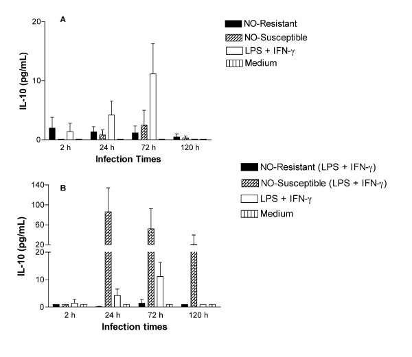
Methods: We evaluated the in vitro toxicity of NO against the promastigotes stages of L. (V.) braziliensis isolated from responsive and refractory patients, and the infectivity of the amastigote forms of these isolates against human macrophages. The supernatants from Leishmania infected macrophage were used to measure TNF-alpha and IL-10 levels.

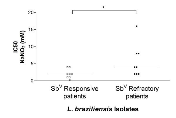
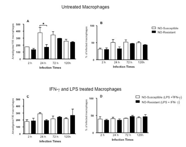
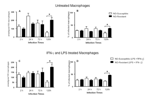
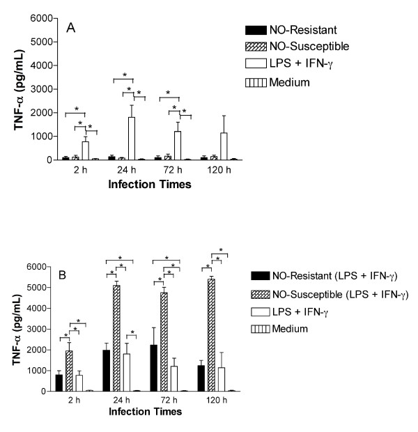
Results: Using NaNO2 (pH 5.0) as the NO source, L. (V.) braziliensis isolated from refractory patients were more NO resistant (IC50 = 5.8 +/- 4.8) than L. (V.) braziliensis isolated from responsive patients (IC50 = 2.0 +/- 1.4). Four isolates were selected to infect human macrophages: NO-susceptible and NO-resistant L. (V.) braziliensis isolated from responsive and refractory patients. NO-resistant L. (V.) braziliensis isolated from refractory patients infected more macrophages stimulated with LPS and IFN-gamma at 120 hours than NO-susceptible L. (V.) braziliensis isolated from refractory patients. Also, lower levels of TNF-alpha were detected in supernatants of macrophages infected with NO-resistant L. (V.) braziliensis as compared to macrophages infected with NO-susceptible L. (V.) braziliensis (p < 0.05 at 2, 24 and 120 hours), while no differences were detected in IL-10 levels.

Conclusion: These data suggest that NO resistance could be related to the nonresponsiveness to antimony therapy seen in American Tegumentary Leishmaniasis.


Figures

Figure 1
Figure 1
IC50 of NaNO2. L. (V.) braziliensis isolated from antimony-refractory patients were more resistant to the NO-generating compound NaNO2 than L. (V.) braziliensis isolated from responsive patients. Eight isolates each from refractory or responsive subject were tested for susceptibility or resistance to NaNO2 using the MTT assay. Lines mean median (* = p = 0.038, Mann-Whitney test). Lb = L. (V.) braziliensis.
Figure 2
Figure 2
Infection of human macrophages with L. (V.) braziliensis (NO-susceptible - open bars and NO-resistant - closed bars) isolated from patients who responded to antimony therapy. A and B, Monocyte-derived macrophages were infected with L. (V.) braziliensis promastigotes (5:1 ratio). At 2, 24, 72 and 120 h of culture(A) the number of intracellular amastigotes and (B) the percentage of infected cells was quantified microscopically as described in Methods. (C) and (D) Monocyte-derived macrophages were treated with LPS (100 ng/mL) plus IFN-γ (10 ng/mL) thirty minutes before infection with L. (V.) braziliensis promastigotes. At 2, 24, 72 and 120 h of culture, (C) the number of intracellular amastigotes and (D) the percentage of infected cells was quantified microscopically. Each bar represents the mean ± SEM infection level in MDMs from three donors (Kruskal-Wallis test, Dunn's multiple comparison Test, * = p < 0.05).
Figure 3
Figure 3
Infection of human macrophages with L. (V.) braziliensis (NO-susceptible - open bars and NO-resistant-closed bars) isolated from patients who were refractory to antimony therapy. (A) and (B) Monocyte-derived macrophages were infected with L. (V.) braziliensis promastigotes (5:1 ratio). At 2, 24, 72 and 120 h of culture the number of intracellular amastigotes(A) and the percentage of infected cells (B) was quantified microscopically as described in Methods. (C) and (D) Monocyte-derived macrophages were treated with LPS (100 ng/mL) plus IFN-γ (10 ng/mL) thirty minutes before the infection with L. (V.) braziliensis promastigotes. At 2, 24, 72 and 120 h of culture, (C) the number of intracellular amastigotes and (D) the percentage of infected cells was quantified microscopically. Each bar represents the mean ± SEM parasite loads in MDMs from three donors (Kruskal-Wallis test, Dunn's multiple comparison Test, * = p < 0.05).
Figure 4
Figure 4
TNF-α production is impaired in macrophages infected with NO-resistant L. (V.) braziliensis. Monocyte-derived macrophages were infected with NO-susceptible or NO-resistant L. (V.) braziliensis. After 2, 24, 72 or 120 h of infection the culture supernatants were harvest from non-stimulated macrophages (A) or macrophages treated with LPS (100 ng/mL) plus IFN-γ (10 ng/mL) thirty minutes prior to the addition of parasites (B). TNF-α levels were determined by ELISA. Each bar represents the mean ± SEM of three experiment from three separate donors (ANOVA, Tukey's multiple comparison Test, * = p < 0.05).
Figure 5
Figure 5
IL-10 production not is altered in macrophages infected with isolated from refractory patients. Monocyte-derived macrophages were infected with NO-susceptible and NO-resistant L. (V.) braziliensis. After 2, 24, 72 or 120 h of infection the culture supernatants of unstimulated macrophages (A) or from macrophages treated with LPS (100 ng/mL) plus IFN-γ (10 ng/mL) for thirty minutes before infection (B), were assessed for IL-10 levels by ELISA. Each bar represents the mean ± SEM infection levels in MDMs from three separate donors.

Similar articles

Cited by

References

    1. Grimaldi G Jr, Tesh RB, McMahon-Pratt D. A review of the geographic distribution and epidemiology of leishmaniasis in the New World. Am J Trop Med Hyg. 1989;41:687–725. - PubMed
    1. Carvalho LP, Passos ST, Jesus AR. Imunopatogênese da Leishmaniose Tegumentar. Gaz Med Bahia. 2005;1:57–65.
    1. Castes M, Trujillo D, Rojas ME, Fernandez CT, Araya L, Cabrera M, Blackwell J, Convit J. Serum levels of tumor necrosis factor in patients with American cutaneous leishmaniasis. Biol Res. 1993;26:233–238. - PubMed
    1. Carvalho EM, Barral A, Costa JM, Bittencourt A, Marsden P. Clinical and immunopathological aspects of disseminated cutaneous leishmaniasis. Acta Trop. 1994;56:315–325. doi: 10.1016/0001-706X(94)90103-1. - DOI - PubMed
    1. Costa JM, Marsden PD, Llanos-Cuentas EA, Netto EM, Carvalho EM, Barral A, Rosa AC, Cuba CC, Magalhaes AV, Barreto AC. Disseminated cutaneous leishmaniasis in a field clinic in Bahia Brazil: a report of eight cases. J Trop Med Hyg. 1986;89:319–323. - PubMed

Publication types

MeSH terms

LinkOut - more resources