

Review
doi: 10.1016/j.brs.2008.09.007.
Epub 2008 Oct 7.
Consensus: New methodologies for brain stimulation
Affiliations
- PMID: 20633398
- PMCID: PMC5507351
- DOI: 10.1016/j.brs.2008.09.007
Item in Clipboard
Review
Consensus: New methodologies for brain stimulation
Brain Stimul.
2009 Jan.
Abstract
We briefly summarized several new stimulation techniques. There are many new methods of human brain stimulation, including modification of already known methods and brand-new methods. In this article, we focused on theta burst stimulation (TBS), repetitive monophasic pulse stimulation, paired- and quadri-pulse stimulation, transcranial alternating current stimulation (tACS), paired associative stimulation, controllable pulse shape TMS (cTMS), and deep-brain TMS. For every method, we summarized the state of the art and discussed issues that remain to be addressed.
Figures

When bursts are given every 200 ms continuously (cTBS), an LTD-like effect induced. On the contrary, when 2-second trains of TBS are given with 8-second breaks in between (iTBS), an LTP-like effect is induced.
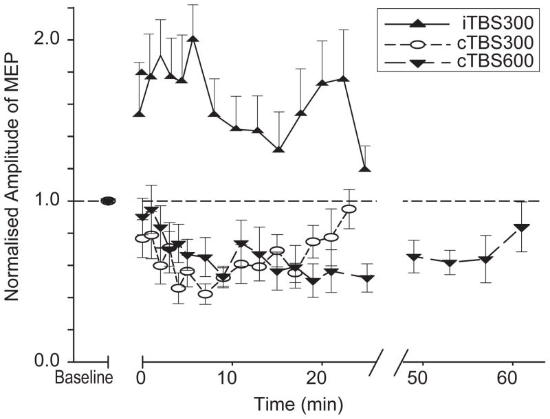
Twenty or 40 seconds of cTBS suppresses the size of MEPs for 20 or 60 minutes, respectively. In contrast, 190 seconds of iTBS enhances MEPs for around 20 minutes.
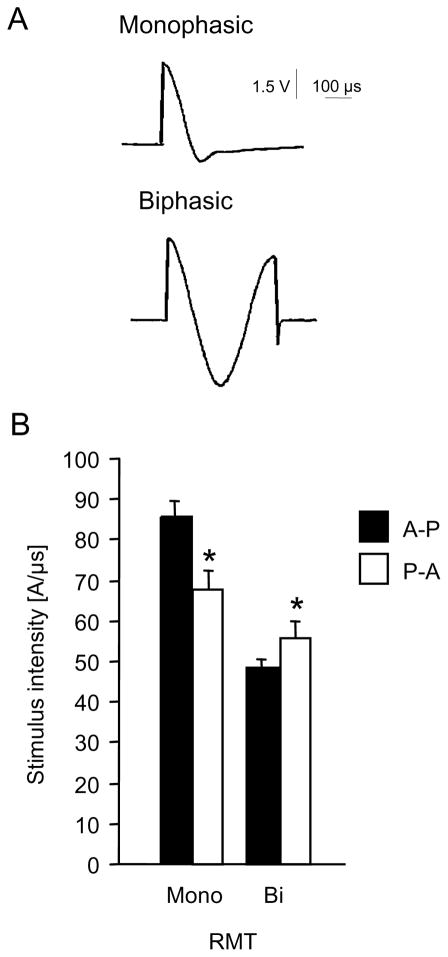
A, current induced in a probe coil of 1 cm diameter by different types of transcranial magnetic stimulators, recorded and stored by an oscilloscope. Upper part, waveform induced by a MagPro stimulator in the “monophasic” mode. Lower part, waveform induced in the “biphasic” mode. B, motor threshold with the target muscle at rest (RMT) in 12 healthy subjects, mean +/− SE. Biphasic (bi) or monophac (mono) stimuli with an anterior (P-A) or posterior (A-P) initial current direction. Asterisks indicate significant post-hoc differences between current directions for a particular waveform. For all graphs the same Dantec MagPro stimulator and the same MC-B70 coil were used. Modified from Sommer et al. 2002 and 2006.
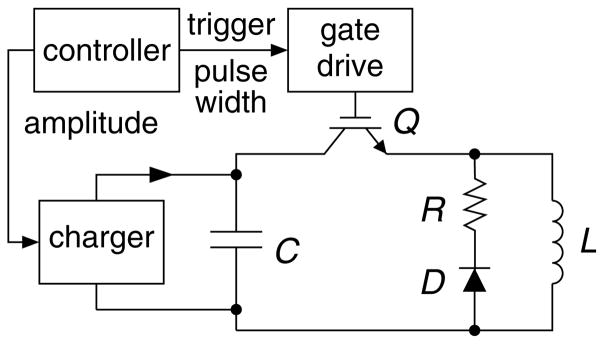
Circuit topology of low-frequency, monophasic cTMS device generating near-rectangular electric field pulses with adjustable pulse width.
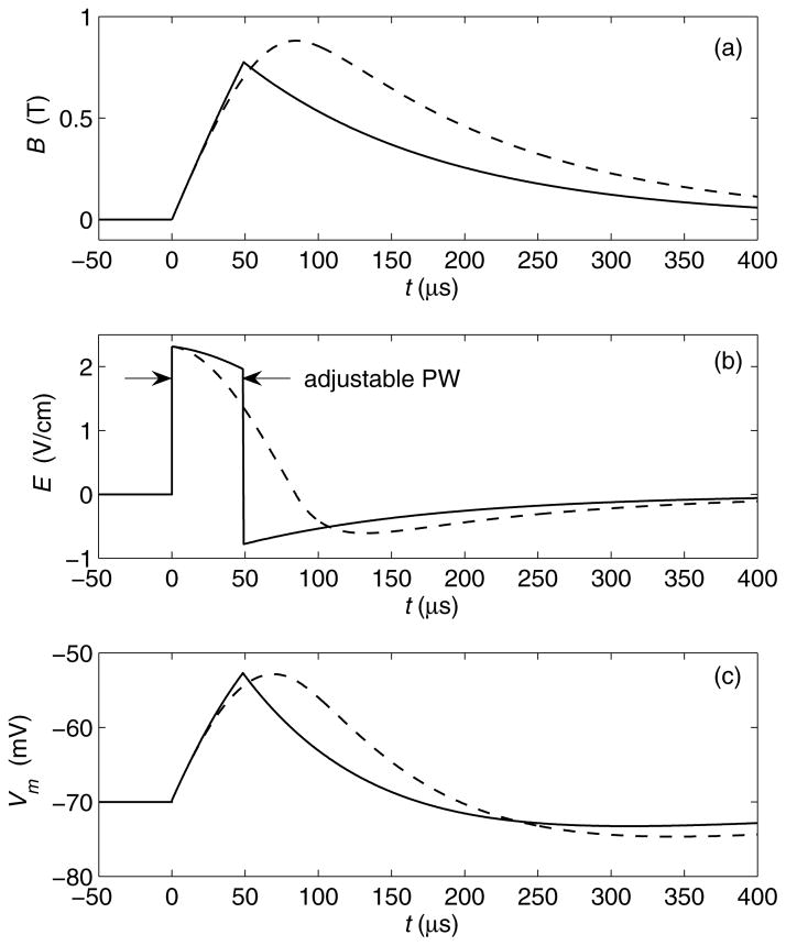
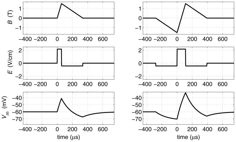
Waveform comparison of monophasic cosine TMS (dashed line) and cTMS (solid line, generated by topology in Fig. 1). (a) Magnetic field B; (b) induced electric field E; (c) neuronal membrane potential Vm for membrane time constant of 150 μs.
Waveforms corresponding to monophasic (left) and biphasic (right) triangular magnetic pulses generated by cTMS device with energy recycling. Same waveform descriptions as in Fig. 5. Energy storage capacitors are assumed to be large.
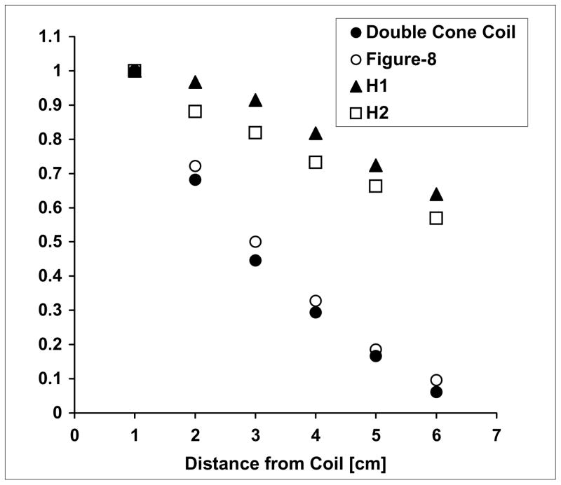
Decay of the electric with distance from various TMS coils. Phantom measurements of the electric field induced at each distance is calculated relative to the field induced 1 cm from the coil, in the ‘z’ (superior-inferior) direction. Data are presented for the H1-coil, H2-coil, double cone coil and figure-8 coil.
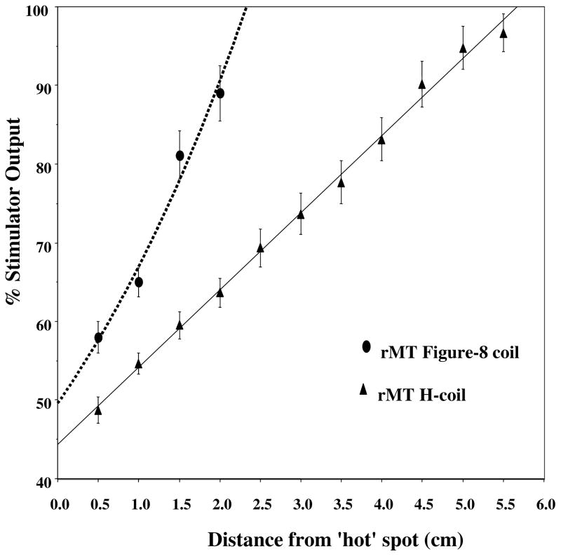
Intensity needed for APB stimulation at different heights above the scalp. Resting motor threshold of the APB was measured at different distances above the ‘hot spot’ when using either the H-coil or the figure-8 coil. The % of stimulator power needed to reach the resting motor threshold vs. the distance of the coil from the ‘hot spot’ on the skull is plotted. The points represent means and standard deviations of 6 healthy volunteers.
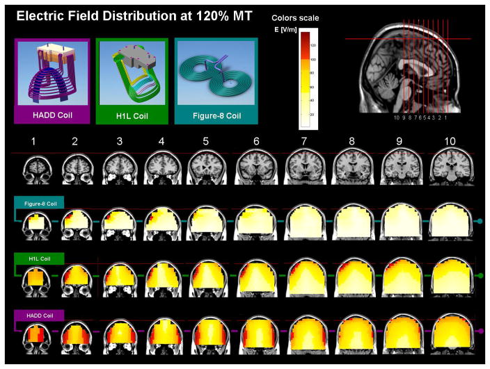
Maps of the electric field induced by three coils placed over the prefrontal cortex. The maps represent phantom brain measurements of the absolute electric field induced at each pixel. The red colors indicate field magnitude above the threshold for neuronal activation, which was set to 100 V/m. The field maps are adjusted for stimulator output required to obtain 120% of the threshold (120 V/m), at a depth of 1.5 cm. a. Field maps of a standard figure-8 coil. b. Field maps of the H1L coil, which was designed to activate lateral prefrontal regions in the left hemisphere. c. Field maps of the HAAD coil, which was designed to activate deep bilateral prefrontal regions.
References
-
- Kandel ER, Spencer WA. Electrophysiology of hippocampal neurons. II. After-potentials and repetitive firing. J Neurophysiol. 1961;24:243–259. - PubMed
-
- Huang YZ, Chen RS, Rothwell JC, Wen HY. The after-effect of human theta burst stimulation is NMDA receptor dependent. Clin Neurophysiol. 2007a;118:1028–1032. - PubMed
-
- Huang YZ, Edwards MJ, Rounis E, Bhatia KP, Rothwell JC. Theta burst stimulation of the human motor cortex. Neuron. 2005;45:201–206. - PubMed
-
- Huang YZ, Rothwell JC. Theta burst stimulation. Transcranial brain stimulation for treatment of psychiatric disorders. In: Marcolin MA, Padberg F, editors. Advances in biological psychiatry. Karger; 2007. pp. 187–203.
-
- Huang YZ, Rothwell JC, Edwards MJ, Chen RS. Effect of Physiological Activity on an NMDA-Dependent Form of Cortical Plasticity in Human. Cereb Cortex. 2007b - PubMed
Publication types
MeSH terms
Grants and funding
LinkOut - more resources
Full Text Sources
Medical
