. 2010 Oct 15;316(17):2797-809.
doi: 10.1016/j.yexcr.2010.07.001. Epub 2010 Jul 13.

A Cajal body-independent pathway for telomerase trafficking in mice

Affiliations

A Cajal body-independent pathway for telomerase trafficking in mice

Rebecca L Tomlinson et al. Exp Cell Res. .

Abstract

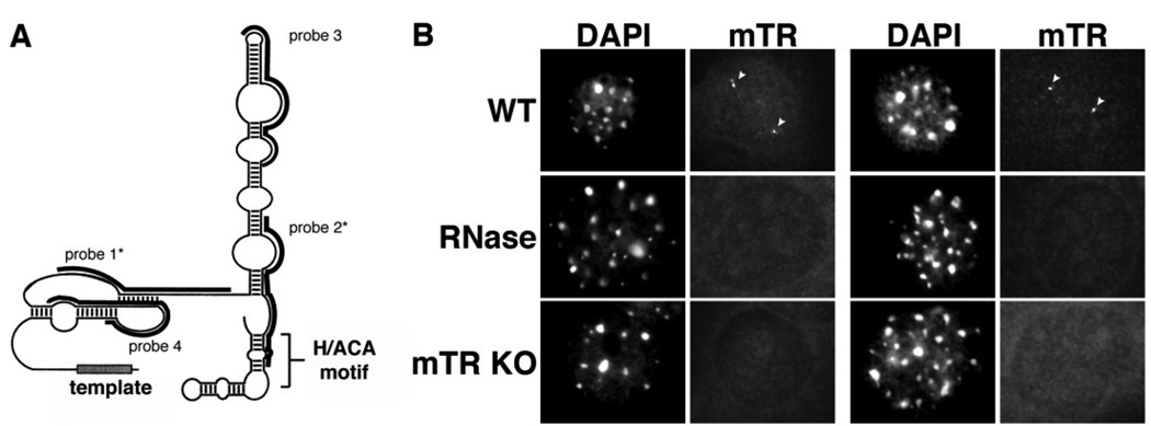
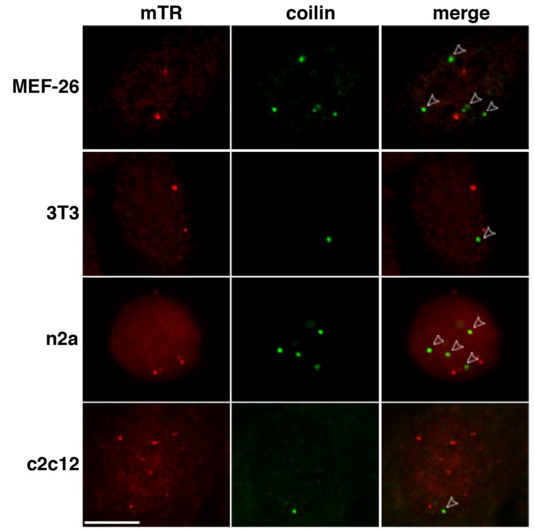
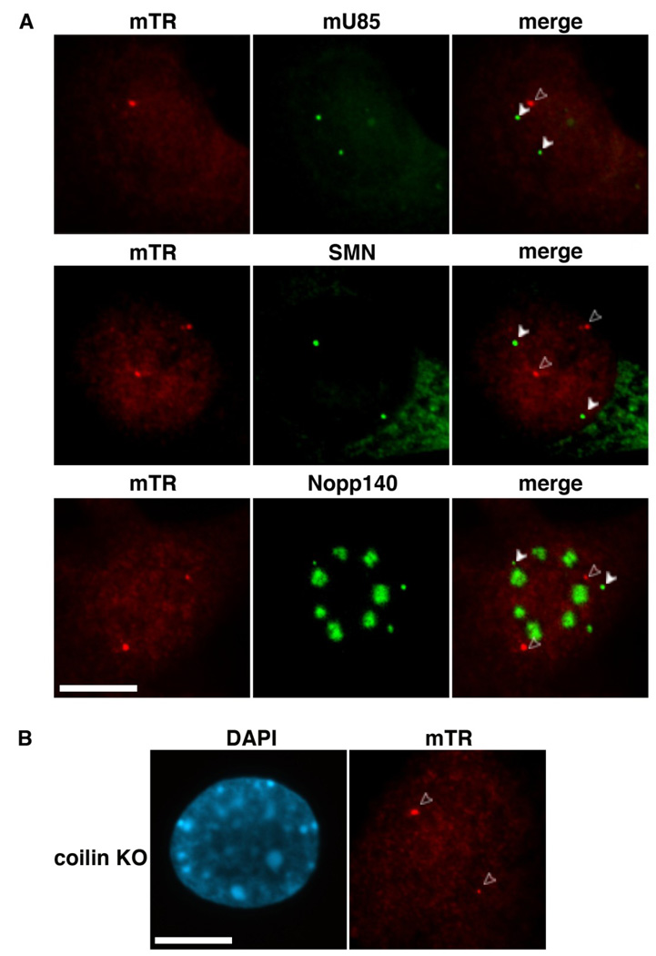
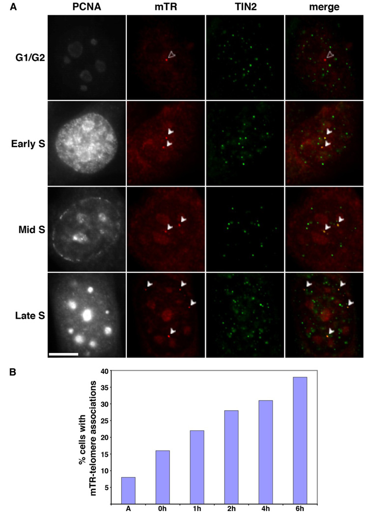
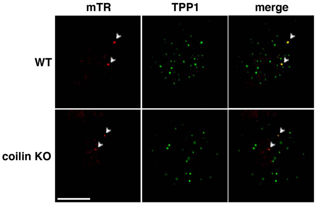
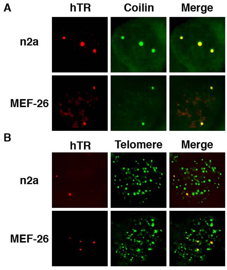
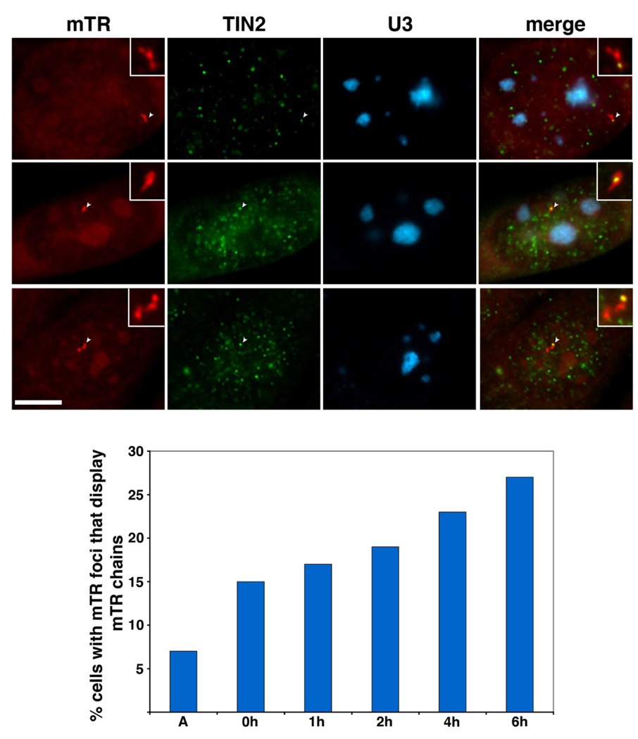
The intranuclear trafficking of human telomerase involves a dynamic interplay between multiple nuclear sites, most notably Cajal bodies and telomeres. Cajal bodies are proposed to serve as sites of telomerase maturation, storage, and assembly, as well as to function in the cell cycle-regulated delivery of telomerase to telomeres in human cells. Here, we find that telomerase RNA does not localize to Cajal bodies in mouse cells, and instead resides in separate nuclear foci throughout much of the cell cycle. However, as in humans, mouse telomerase RNA (mTR) localizes to subsets of telomeres specifically during S phase. The localization of mTR to telomeres in mouse cells does not require coilin-containing Cajal bodies, as mTR is found at telomeres at similar frequencies in cells from wild-type and coilin knockout mice. At the same time, we find that human TR localizes to Cajal bodies (as well as telomeres) in mouse cells, indicating that the distinct trafficking of mTR is attributable to an intrinsic property of the RNA (rather than a difference in the mouse cell environment such as the properties of mouse Cajal bodies). We also find that during S phase, mTR foci coalesce into short chains, with at least one of the conjoined mTR foci co-localizing with a telomere. These findings point to a novel, Cajal body-independent pathway for telomerase biogenesis and trafficking in mice.


Figures

Figure 1
Figure 1. Mouse telomerase RNA is found in small spherical foci within the nuclei of cultured mouse cell lines
A. Schematic structure of mTR. The predicted secondary structure of mTR is shown [70]. Black bars indicate the regions encompassed by each oligonucleotide probe. Asterisks denote the two probes (probes 1 and 2) used throughout this manuscript. B. FISH procedure specifically detects the presence of mTR. mTR FISH was performed on wild type MEF cells (WT, RNAse panels) or MEF cells derived from mTR −/− mice (mTR KO panels). Arrowheads denote intranuclear mTR foci present in the WT cells (WT panels), which are lost upon treatment of cells with RNAse A prior to FISH (RNAse panels). DAPI was used as a nuclear stain. Scale bar, 10 microns.
Figure 2
Figure 2. mTR foci do not colocalize with coilin in a variety of mouse cell lines
Mouse embryonic fibroblast (MEF-26, 3T3), n2a neuroblastoma, and c2c12 myoblast cell lines were co-analyzed for mTR (detected by FISH, red) and coilin (marker protein for Cajal bodies, detected by IF, green). Merge panels indicate an overlay of mTR and coilin panels. Open arrowheads point to Cajal bodies that do not overlap with mTR foci. Scale bar, 10 microns.
Figure 3
Figure 3. mTR resides in foci separate from Cajal bodies
A. mTR foci do not correspond to known markers for Cajal bodies. mTR FISH (red, mTR panels) was performed in tandem with one of three markers for Cajal bodies: U85 scaRNA (top row, detected by FISH), SMN (middle row, detected by IF, signal present in Cajal bodies and cytoplasm), or Nopp140 (bottom row, detected by IF, signal present in Cajal bodies and nucleoli). Arrowheads denote Cajal bodies; open arrowheads point to mTR foci that do not localize to Cajal bodies. B. mTR localizes to intranuclear foci in MEF cells derived from coilin knockout (KO) mice. mTR FISH was performed on coilin KO MEF cells. Open arrowheads point to mTR foci. DAPI was used to stain the DNA. Scale bars, 10 microns.
Figure 4
Figure 4. mTR localizes to subsets of telomeres in S phase
A. Localization of mTR to telomeres occurs throughout S phase. mTR FISH (red) and TIN2 IF (green, marker for telomeres) were performed on 3T3 cells at various stages of the cell cycle. PCNA staining was performed to identify S phase cells, as well as S sub-phase (i.e. early, mid, or, late, as indicated). Merge panels show a superimposition of mTR and TIN2 panels, yellow indicates an overlap of signal. The foci where mTR and telomeres colocalize are indicated by white arrowheads. Open arrowheads (in G1/G2 panel) denotes mTR foci that do not overlap with a telomere. Scale bar, 10 microns. B. The frequency of mTR-telomere associations increases as cells progress through S phase. The percentage of 3T3 cells with mTR foci that display mTR at the telomere (assessed via TIN2) is plotted relative to time in hours (h) after release from a double thymidine block. A, asynchronous cells. Data was collected from multiple slides prepared as part of at least two separate experiments.
Figure 5
Figure 5. mTR localizes to the telomere in coilin KO MEFs
mTR FISH (red) and TPP1 IF (green, marker for telomeres) were performed on MEF cells derived from wild type (WT) and coilin −/− (coilin KO) MEFs. Merge panels display an overlay of mTR and TPP1 signals. Arrowheads denote foci where both mTR and TPP1 signals overlap. Scale bar, 10 microns.
Figure 6
Figure 6. Human telomerase RNA localizes to Cajal bodies and telomeres in mouse cells
A. hTR co-localizes with Cajal bodies in mouse cells. Human telomerase RNA (hTR) was expressed in n2a and MEF-26 mouse cell lines and co-analyzed for hTR (detected by FISH, red panels) and coilin (detected by IF, green panels). Merge panels display an overlay of hTR and coilin signals. B. hTR co-localizes with telomeres in mouse cells. hTR-transfected n2a and MEF-26 mouse cells were examined for hTR (detected by FISH, red panels) and telomere (detected by FISH, green panels) signals. Merge panels display an overlay of hTR and telomere signals.
Figure 7
Figure 7. mTR foci coalesce during S phase of the cell cycle
A. mTR foci link together to form chains that are frequently associated with telomeres but not nucleoli. 3T3 cells were stained for mTR (detected by FISH, red panels), TIN2 (telomere marker detected by IF, green panels), and U3 snoRNA (nucleolar marker detected by FISH, blue panels). Merge panels display a superimposition of all 3 panels. Insets show an enhanced magnification of the mTR chains. Arrowheads point to mTR foci within the chain that overlap with a telomere. Scale bar, 10 microns. B. The frequency of mTR chains gradually increases over S phase of the cell cycle. The percentage of 3T3 cells with mTR foci that display a mTR chain are plotted relative to time after release from a double thymidine block. A, asynchronous cells. Data compiled from two separate experiments.

References

    1. de Lange T. Shelterin: the protein complex that shapes and safeguards human telomeres. Genes Dev. 2005;19:2100–2110. - PubMed
    1. Xin H, Liu D, Songyang Z. The telosome/shelterin complex and its functions. Genome Biol. 2008;9:232. - PMC - PubMed
    1. Palm W, de Lange T. How shelterin protects mammalian telomeres. Annu Rev Genet. 2008;42:301–334. - PubMed
    1. Armanios M. Syndromes of Telomere Shortening. Annu Rev Genomics Hum Genet. 2009 - PMC - PubMed
    1. Artandi SE. Telomeres, telomerase, and human disease. N Engl J Med. 2006;355:1195–1197. - PubMed

Publication types