. 2010 Oct;137(1):74-80.
doi: 10.1016/j.clim.2010.06.011. Epub 2010 Jul 14.

TLR signaling and effector functions are intact in XLA neutrophils

Affiliations

TLR signaling and effector functions are intact in XLA neutrophils

Thomas U Marron et al. Clin Immunol. 2010 Oct.

Abstract

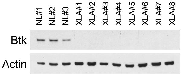
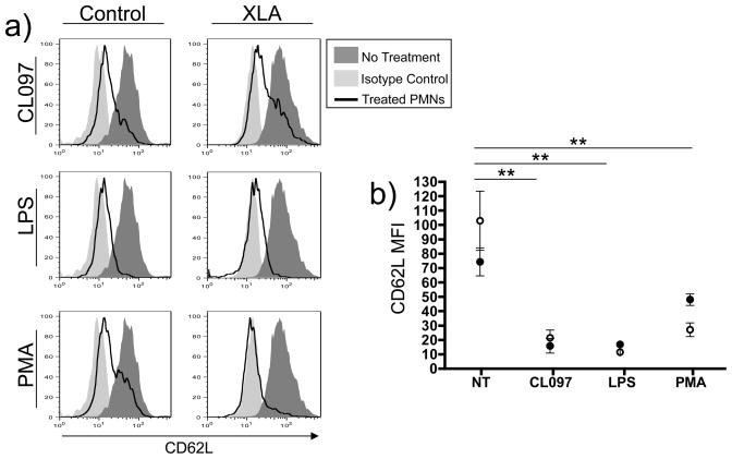
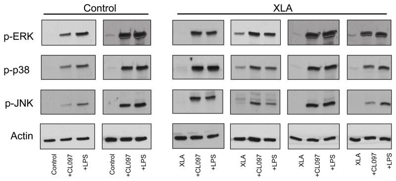
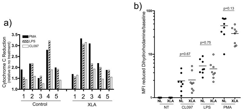
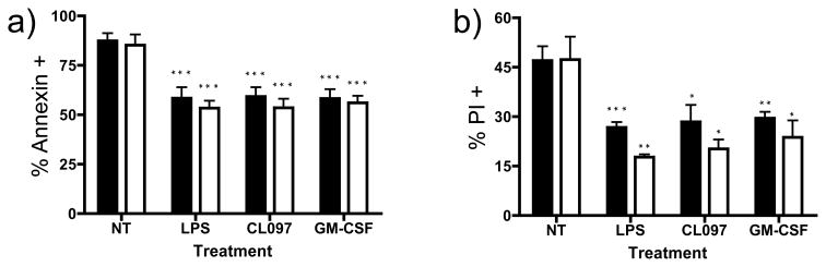
Toll-like receptors (TLRs) are essential components of the innate immune system, and their ligands are important activators of neutrophils. Bruton's tyrosine kinase (Btk) has been reported to mediate signaling through toll-like receptors (TLRs) in many cell types, however, the role of Btk in TLR activation of neutrophils remains unclear. Impaired TLR-induced neutrophil function was found in mice with loss of Btk and in humans with TLR-signaling defects, but the integrity of TLR pathways in X-linked agammaglobulinemia (XLA) neutrophils has not been assessed. In this study LPS (TLR4) or an imidazoquinoline compound (TLR7/8) activated XLA neutrophil shedding of surface CD62L, and phosphorylated MAP kinases p38, JNK and ERK. TLR activation also induced normal respiratory burst and retarded apoptosis for XLA neutrophils, comparable to normal controls. These data demonstrate that the loss of Btk in XLA neutrophils does not impair functional responses to TLR signals.


Figures

Figure 1
Figure 1. Btk expression in XLA cells
Leukocytes of 8 XLA patients examined by western blot, demonstrated no Btk expression in contrast to cells of 3 normal controls.
Figure 2
Figure 2. Neutrophil CD62L shedding
Whole blood was treated with TLR ligands CL097 (2.5μg/mL) or LPS 100ng/mL or PMA 10ng/mL for 1 hour, followed by RBC lysis and analysis of CD62L shedding from the surface of neutrophils by FACS. Representative histograms of gated granulocytes from a control and one XLA subject, demonstrated loss of CD62L with TLR activation (a). The MFI of FACS analyses from XLA subjects (open circles, n=6) and controls (solid circles, n=6) is represented graphically (b). An unpaired two-tailed t-test used to individually compare CD62L expression on untreated XLA cells with treatment groups. ** denotes a p value of <0.005.
Figure 3
Figure 3. MAPK signaling
Neutrophils isolated from blood of controls (n=6) or XLA subjects (n=6) were treated for 5 minutes with CL097 (2.5μg/mL) or LPS (100ng/mL), and phosphorylation of MAP kinases p38, JNK and ERK was assessed by western blot. Representative results for two controls and four XLA subject are shown in comparison to β-actin.
Figure 4
Figure 4. Figure 4a and 4b: TLR induction of respiratory burst
Isolated neutrophils from normal controls (n=5) or XLA subjects (n=5) were first incubated with cytochrome C, in the presence or absence of SOD, and treated for 20 minutes with CL097 (2.5μg/mL), LPS (100ng/mL) or PMA (10ng/mL). (a) Reduction of cytochrome C by the superoxide radicals produced by neutrophils was assessed at 550nM absorbance. (b) Isolated neutrophils from normal controls (solid circles, n=8) and XLA subjects (open circles, n=8) were incubated with dihydrorhodamine and treated as in Figure 4a, and reduction of dihydrorhodamine to rhodamine was assessed by FACS. These results are represented graphically as MFI of the rhodamine signal. p-values shown are from a two-tailed t-test comparing control and XLA responses within each treatment group.
Figure 5
Figure 5. Assessing neutrophil apoptosis
Isolated neutrophils from normal controls (solid bars, n=4) or XLA subjects (open bars, n=4) were cultured for 48 hours with CL097 (2.5μg/mL), LPS (100ng/mL) or GM-CSF (20ng/mL), and induction of apoptosis assessed by Annexin V, (5a) or late apoptosis PI, (5b). Data is represented as a percentage of Annexin or PI positive cells. P values denoted represent two-tailed t-test comparison of each treatment to the untreated control neutrophils. No differences were seen between normal and XLA neutrophils in any treatment group. Noted significance (*p<0.05, **p<0.01, ***p<0.005) represents comparison to untreated neutrophils from same patient or control subject.

References

    1. Conley ME. B cells in patients with X-linked agammaglobulinemia. Journal of Immunology. 1985;134:3070–3074. - PubMed
    1. Vetrie D, Vorechovsky I, Sideras P, Holland J, Davies A, Flinter F, Hammarstrom L, Kinnon C, Levinsky R, Bobrow M. The gene involved in X-linked agammaglobulinaemia is a member of the src family of protein-tyrosine kinases. Nature. 1993;361:226–233. [see comment][erratum appears in Nature 1993 Jul 22;364(6435):362] - PubMed
    1. Conley ME. Genetics of hypogammaglobulinemia: what do we really know? Curr Opin Immunol. 2009;21:466–471. - PMC - PubMed
    1. Farrar JE, Rohrer J, Conley ME. Neutropenia in X-linked agammaglobulinemia. Clinical Immunology & Immunopathology. 1996;81:271–276. - PubMed
    1. Winkelstein JA, Marino MC, Lederman HM, Jones SM, Sullivan K, Burks AW, Conley ME, Cunningham-Rundles C, Ochs HD. X-linked agammaglobulinemia: report on a United States registry of 201 patients. Medicine. 2006;85:193–202. - PubMed

Publication types

MeSH terms