Comparative Study
. 2010 Jul 15;24(14):1533-45.
doi: 10.1101/gad.1936910.

Dissecting the functional specificities of two Hox proteins

Affiliations
Comparative Study

Dissecting the functional specificities of two Hox proteins

Rohit Joshi et al. Genes Dev. .

Abstract

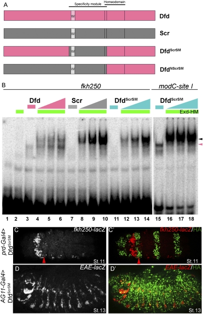
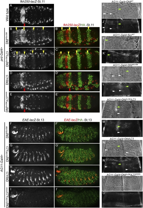
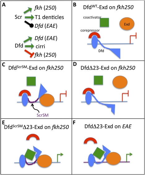
Hox proteins frequently select and regulate their specific target genes with the help of cofactors like Extradenticle (Exd) and Homothorax (Hth). For the Drosophila Hox protein Sex combs reduced (Scr), Exd has been shown to position a normally unstructured portion of Scr so that two basic amino acid side chains can insert into the minor groove of an Scr-specific DNA-binding site. Here we provide evidence that another Drosophila Hox protein, Deformed (Dfd), uses a very similar mechanism to achieve specificity in vivo, thus generalizing this mechanism. Furthermore, we show that subtle differences in the way Dfd and Scr recognize their specific binding sites, in conjunction with non-DNA-binding domains, influence whether the target gene is transcriptionally activated or repressed. These results suggest that the interaction between these DNA-binding proteins and the DNA-binding site determines the architecture of the Hox-cofactor-DNA ternary complex, which in turn determines whether the complex recruits coactivators or corepressors.


Figures

Figure 1.
Figure 1.
Analysis of modC from Dfd. (A) Schematic of 2.7-kb EAE-lacZ and 570-bp modC-lacZ. Sequence of Dfd-, Exd-, and Hth-binding sites in modC are color-coded. Mutant versions of modC are shown below the wild-type sequence. The Dfd–Exd composite binding site (site I) and nearby Hth-binding site is labeled “I.” Other combinations of Dfd-, Exd-, and Hth-binding sites are indicated as II, III, and IV. Note that Exd-binding sites often overlap with Hth-binding sites. (B,C) Epidermal expression of EAE-lacZ (B) and modC-lacZ (C) in maxillary and mandibular segments of the embryo. In these and all subsequent images, costaining for Scr in the right panels is used to identify the adjacent Dfd-expressing segments. Anterior is always to the left, and all embryos are at stage 13. (D,E) Mutagenesis of Dfd–Exd (D) or Exd half-site (E) of site I in modC results in a modest decrease in reporter gene activity (DE-lacZ and E-lacZ, respectively). In these and subsequent images, the yellow arrowheads point to the maxillary segments. (F) Mutagenesis of four Dfd-binding sites in the modC enhancer significantly decreases enhancer activity (4D-lacZ). (G) Mutagenesis of the Dfd–Exd site from site I and four Dfd-binding sites abolishes enhancer activity (DE-4D-lacZ). (H) Mutagenesis of the Exd half-site from site I and four Dfd-binding sites abolishes enhancer activity (E4D-lacZ). (I) modC-lacZ is induced in response to ectopic Dfd by AG11-Gal4.
Figure 2.
Figure 2.
Dfd-specific functions depend on Exd–Hth. (A,B) modC-lacZ expression is normal in the maxillary segment (yellow arrowheads) of hthP2 heterozygotes (A; visualized by hb-lacZ marked balancer), but is abolished in hthP2 homozygotes (B). The ectopic expression driven by modC in hthP2 embryos suggests that Hth may have a repressive role in thoracic and abdominal segments. (C) modC-lacZ requires direct Exd–Hth input. Mutagenesis of three Exd–Hth-binding sites in modC (3EH-lacZ) (Fig. 1A) results in a large decrease in enhancer activity. Note that Dfd monomer binding to this element is weakly affected by this mutation. (D) Hth binds to modC in vivo. Anti-Hth antibody was used to precipitate embryonic chromatin. Immunoprecipitated chromatin was assayed for the presence of modC (spanning site I) and a negative control locus, PDH. Data are presented as a percentage of the signal obtained relative to input chromatin and represent the averages and standard deviation of two independent ChIPs. (E, lanes 7,8) DfdWT binds cooperatively to site I with Exd–HM. (Lanes 4,5) DfdYPAA fails to bind cooperatively with Exd–HM to site I. (Lanes 12,13) DfdWT fails to bind cooperatively with Exd–HM to site I that has a mutant Exd half-site (site Iexd-mut). Hox monomer and Hox–Exd–HM trimer complexes are indicated by the pink and black arrowheads, respectively. (F,G) Ectopic expression of DfdWT (F) results in ectopic activation of EAE-lacZ, while ectopic expression of DfdYPAA (G) does not (see Fig. 3F for quantification). (H) Head and thorax of a wild-type embryo showing the normal position of cirri in the head segment (indicated by pink arrowhead). Ubiquitous expression of DfdWT (H′) results in ectopic cirri (yellow arrowheads, see inset in panel) and mouth hooks in thoracic segments (see Fig. 3G for quantification), but expression of DfdYPAA failed to induce any Dfd-specific structures (H″).
Figure 3.
Figure 3.
Dfd specificity module residues His-15 and Arg3 are required in vitro and in vivo for Dfd-specific functions. (A) Schematic showing Hox specificity module, homeodomain, YPWM motif, and linker. Below are alignments of specificity modules from Dfd orthologs, showing the conserved histidine and an arginine (underlined) at positions analogous to those in Scr's specificity module (Joshi et al. 2007). (B) DfdHis-15A, Arg3A binding to site I is weaker both as a monomer (lane 6) and as cooperative trimer (lanes 7,8) with Exd–HM in comparison with DfdWT (lanes 35). Hox monomer and Hox–Exd–HM trimer complexes are indicated by pink and black arrowheads, respectively. (C) Kd measurements for DfdWT–Exd–HM and DfdHis-15A, Arg3A–Exd–HM to site I show an eightfold difference in binding affinity. (D,E) Ubiquitous expression of DfdWT results in ectopic activation of EAE-lacZ (D), while DfdHis-15A, Arg3A (E) is severely compromised in its ability to induce this reporter gene (see F for quantification). (F) Graph showing percentage of total embryos that activate EAE-lacZ ectopically when DfdWT, DfdHis-15A, Arg3A, or DfdYPAA is expressed ubiquitously using AG11-Gal4 (n ≥ 56 for each genotype, n = 2). (G) Graph showing the quantification of ectopic cirri made in thoracic segments of embryonic cuticles when DfdWT or DfdHis-15A, Arg3A is expressed ubiquitously using AG11-Gal4. Error bars represent standard deviations.
Figure 4.
Figure 4.
Dfd represses fkh250-lacZ in an Exd-independent manner. (A) Wild-type expression patterns of fkh250-lacZ and prd-GAL4 (driving GFP) to show overlap between these two expression domains. The strongest expression of fkh250-lacZ is in PS2. (B) Ectopic expression of ScrWT activates fkh250-lacZ (yellow arrowheads). (C) Ectopic expression of DfdWT represses fkh250-lacZ (red arrowhead). (D) Ectopic expression of DfdHis-15A, Arg3A represses fkh250-lacZ (red arrowhead), showing that repression of fkh250-lacZ by Dfd is independent of these residues. (E) Ectopic expression of DfdAAAA represses fkh250-lacZ (red arrowhead), suggesting that repression of fkh250-lacZ by Dfd does not require recruitment of Exd by its YPWM motif. (F) Coexpression of both DfdWT and ScrWT represses fkh250-lacZ in PS2 and largely blocks its activation in posterior segments (white arrowheads), showing that fkh250-lacZ repression by DfdWT is direct. (G) Coexpression of both DfdAAAA and ScrWT represses fkh250-lacZ in PS2 and largely blocks its activation in posterior segments (white arrowheads), showing that repression of fkh250-lacZ by Dfd is direct and unlikely to require Exd recruitment.
Figure 5.
Figure 5.
Enhancing Exd recruitment by Dfd to fkh250 is not sufficient for activation. (A) Schematics of DfdScrSM and DfdNScrSM chimeras. Extent of specificity modules and homeodomains are indicated (for details, see the text). (B) DfdWT binds less cooperatively with Exd–HM to fkh250 (lanes 46) compared with ScrWT (lanes 810). (Lanes 1214) DfdScrSM binds very cooperatively with Exd–HM to fkh250. (Lanes 1618) DfdScrSM binds very cooperatively with Exd–HM on site I. Hox monomer and Hox–Exd–HM trimer complexes are indicated by pink and black arrowheads, respectively. (C,D) Ectopic expression of DfdScrSM represses fkh250-lacZ (C; red arrowhead) and activates EAE-lacZ (D), suggesting that an enhancement of Exd recruitment is not sufficient for fkh250-lacZ activation.
Figure 6.
Figure 6.
Changing the functional specificity of Dfd. (A) Wild-type expression pattern of fkh250-lacZ. (B,C) Ectopic expression of DfdNScrSM (B) or DfdScrSMΔ23 (C) activates fkh250-lacZ (yellow arrowheads), showing a change in specificity. (D,E) Ectopic expression of DfdΔ23 (D) or DfdScrSMΔ23AAAA (E) represses fkh250-lacZ in PS2 (red arrowhead), emphasizing the importance of Scr's SM- and Exd-mediated cooperative binding in activation of fkh250-lacZ. (FH) Ectopic expression of DfdNScrSM (F), DfdScrSMΔ23 (G), or DfdΔ23 (H) activates EAE-lacZ. These proteins retain the ability to activate Dfd, like DfdWT. (I) Ectopic expression of DfdScrSMΔ23AAAA does not activate EAE-lacZ, emphasizing the importance of Exd recruitment in EAE-lacZ activation. (JO) Cuticle phenotypes resulting from ubiquitous expression of DfdWT (J), ScrWT (K), DfdNScrSM (L), DfdScrSMΔ23 (M), DfdΔ23 (N), and DfdScrSMΔ23AAAA (O). Scr-specific structures (ectopic T1 beard) are indicated with green arrowheads, and Dfd-specific structures (cirri) are indicated by white arrowheads. Dark-field and phase-contrast images for the T2 segment are shown for all cuticles.
Figure 7.
Figure 7.
Interplay between DNA-binding specificity and transcriptional output. (A) Specific in vivo functions of Scr and Dfd. Green arrows indicate specific functions, the repression bar is indicated in red, and the black line indicates no effect. (BF) Schematized is a subset of the various regulatory scenarios examined in this study. Activation is indicated by a green arrow, and the repression bar is indicated in red. A comparison between DfdScrSM–Exd bound to fkh250 (C), which represses transcription, and DfdScrSMΔ23–Exd bound to fkh250 (E), which activates transcription, suggests that motifs 2 and 3 are required to either recruit a corepressor or block the recruitment of a coactivator. A comparison between DfdΔ23–Exd bound to fkh250 (D), which represses transcription, and DfdScrSMΔ23–Exd bound to fkh250 (E), which activates transcription, suggests that differences in the specificity module, and thus how these proteins bind to fkh250, determines the sign of the regulation. Finally, a comparison between DfdΔ23–Exd bound to fkh250 (D), which represses transcription, and DfdΔ23–Exd bound to EAE (F), which activates transcription, suggests that differences between binding sites also contribute to differences in regulatory output.

Similar articles

Cited by

References

    1. Abu-Shaar M, Ryoo HD, Mann RS 1999. Control of the nuclear localization of Extradenticle by competing nuclear import and export signals. Genes Dev 13: 935–945 - PMC - PubMed
    1. Agelopoulos M, Thanos D 2006. Epigenetic determination of a cell-specific gene expression program by ATF-2 and the histone variant macroH2A. EMBO J 25: 4843–4853 - PMC - PubMed
    1. Berger MF, Badis G, Gehrke AR, Talukder S, Philippakis AA, Pena-Castillo L, Alleyne TM, Mnaimneh S, Botvinnik OB, Chan ET, et al. 2008. Variation in homeodomain DNA binding revealed by high-resolution analysis of sequence preferences. Cell 133: 1266–1276 - PMC - PubMed
    1. Bergson C, McGinnis W 1990. An autoregulatory enhancer element of the Drosophila homeotic gene Deformed. EMBO J 9: 4287–4297 - PMC - PubMed
    1. Bischof J, Maeda RK, Hediger M, Karch F, Basler K 2007. An optimized transgenesis system for Drosophila using germ-line-specific phiC31 integrases. Proc Natl Acad Sci 104: 3312–3317 - PMC - PubMed

Publication types

MeSH terms

LinkOut - more resources