. 2010 Jul 15;24(14):1546-58.
doi: 10.1101/gad.573310.

Loss of Topoisomerase I leads to R-loop-mediated transcriptional blocks during ribosomal RNA synthesis

Affiliations

Loss of Topoisomerase I leads to R-loop-mediated transcriptional blocks during ribosomal RNA synthesis

Aziz El Hage et al. Genes Dev. .

Abstract

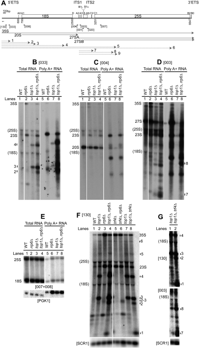
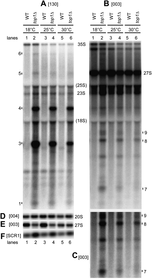
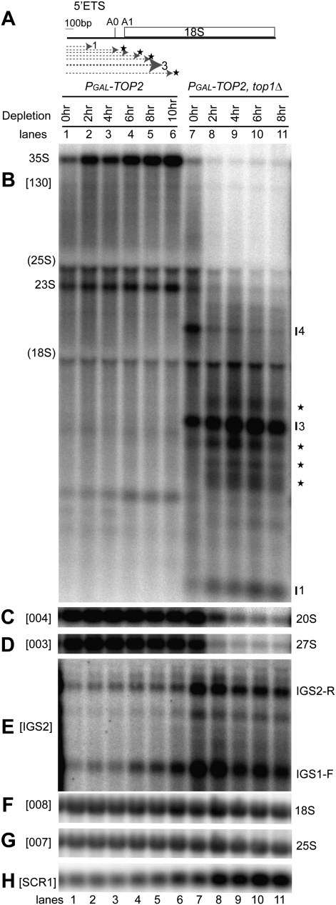
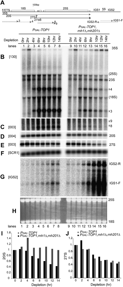
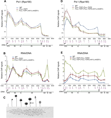
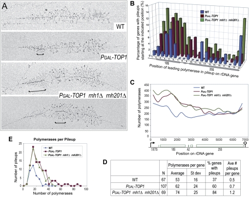
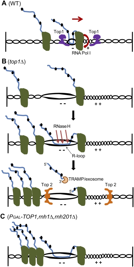
Pre-rRNA transcription by RNA Polymerase I (Pol I) is very robust on active rDNA repeats. Loss of yeast Topoisomerase I (Top1) generated truncated pre-rRNA fragments, which were stabilized in strains lacking TRAMP (Trf4/Trf5-Air1/Air2-Mtr4 polyadenylation complexes) or exosome degradation activities. Loss of both Top1 and Top2 blocked pre-rRNA synthesis, with pre-rRNAs truncated predominately in the 18S 5' region. Positive supercoils in front of Pol I are predicted to slow elongation, while rDNA opening in its wake might cause R-loop formation. Chromatin immunoprecipitation analysis showed substantial levels of RNA/DNA hybrids in the wild type, particularly over the 18S 5' region. The absence of RNase H1 and H2 in cells depleted of Top1 increased the accumulation of RNA/DNA hybrids and reduced pre-rRNA truncation and pre-rRNA synthesis. Hybrid accumulation over the rDNA was greatly exacerbated when Top1, Top2, and RNase H were all absent. Electron microscopy (EM) analysis revealed Pol I pileups in the wild type, particularly over the 18S. Pileups were longer and more frequent in the absence of Top1, and their frequency was exacerbated when RNase H activity was also lacking. We conclude that the loss of Top1 enhances inherent R-loop formation, particularly over the 5' region of the rDNA, imposing persistent transcription blocks when RNase H is limiting.


Figures

Figure 1.
Figure 1.
Aberrant pre-rRNAs generated in the absence of Top1 are targeted for degradation by TRAMP and exosome complexes. (A) Schematic representation of a 35S rRNA gene in S. cerevisiae. Pre-rRNA processing sites A0, A1, D, A2, A3, B1, C2, C1, B2, and B0 are labeled. Probes used in Northern analyses are indicated by bars. The 35S, 20S, 27SA, and 27SB pre-rRNAs are depicted by solid arrows. Truncated pre-rRNA fragments generated in the absence of Top1 are depicted by dotted arrows and are labeled 1–9. Numbers of nucleotides along the rDNA unit relative to the start of transcription (0) are indicated. The pre-rRNA processing pathway is depicted in Supplemental Figure S1. (B–E) Polyadenylated pre-rRNA fragments accumulate in double top1Δ rrp6Δ mutants. Total RNA was extracted from wild-type (WT), rrp6Δ, top1Δ, and top1Δ rrp6Δ strains grown at 25°C. Polyadenylated RNAs were purified and resolved alongside total RNAs on a 1.2% glyoxal-agarose gel. Pre-rRNA fragments were detected by Northern analysis: probe 033, hybridizing at +278 in 5′-ETS (B); probe 004, hybridizing upstream of site A2 (C); and probe 003, hybridizing between A2 and A3 (D). (E) As controls for poly(A)+ RNA specificity, 25S and 18S rRNA and PGK1 mRNA were analyzed. (F,G) TRAMP participates in pre-rRNA surveillance in top1Δ mutants. Total RNA was extracted from wild-type and single and double top1Δ, trf4Δ, and rrp6Δ strains grown at 25°C and analyzed using probe 130 (hybridizes in the 5′-ETS upstream of probe 033) and 003. F and G are from two independent experiments. (Bottom panels) SCR1 was used as a loading control. Intact pre-rRNAs and truncated pre-rRNA fragments are labeled. “(18S)” and “(25S)” indicate the positions of migration of 18S and 25S rRNAs. Probe names are bracketed. The 23S RNA is an aberrant pre-rRNA processing intermediate extending from +1 to site A3, shown previously to be polyadenylated and targeted for degradation by TRAMP and Rrp6 (Dez et al. 2006).
Figure 2.
Figure 2.
Pre-rRNA synthesis defects increase in top1Δ mutant at low growth temperatures. Total RNA from wild-type and single top1Δ strains grown at 18°C, 25°C, or 30°C was resolved on an agarose gel and analyzed by Northern hybridization, using probes 130 (A), 003 (B,C,E), and 004 (D). (F) Cytoplasmic SCR1 RNA was used as a loading control. C shows a stronger exposure of the lower region (below 18S) of B. Intact pre-rRNAs and truncated pre-rRNA fragments are labeled. “(18S)” and “(25S)” indicate the position of migration of 18S and 25S rRNAs. Probe names are bracketed (see Fig. 1A for location of probes).
Figure 3.
Figure 3.
Pre-rRNAs are truncated predominately within the 5′ region of 18S rRNA when both Top1 and Top2 are absent. (A) Diagram representing the 5′-ETS and 18S rDNA sequences. Heterogeneous, truncated pre-rRNA fragments that accumulate in the absence of both Top1 and Top2 are depicted by dotted arrows followed by a star. (B–H) Strains PGAL-TOP2 and PGAL-TOP2 top1Δ were shifted from galactose-containing medium (0 h) to glucose-containing medium at 30°C (2–10 h). Total RNA was analyzed by Northern hybridization. The membrane was hybridized successively with probes 130 (B), 004 (C), 003 (D), 008 (F), and 007 (G). (E) Random primed probe IGS2 was used to detect ncRNAs transcribed by RNA Pol II from the intergenic rDNA spacers IGS1 and IGS2, located between the 35S rDNA units. (H) Cytoplasmic SCR1 RNA was used as a loading control. Intact pre-rRNAs and truncated pre-rRNA fragments are labeled. Heterogeneous truncated pre-rRNA fragments are labeled by stars. “(18S)” and “(25S)” indicate the position of migration of 18S and 25S rRNAs. Probe names are bracketed (see Fig. 1A for location of probes).
Figure 4.
Figure 4.
RNase H facilitates pre-rRNA transcription in the absence of Top1. (A) Schematic showing the locations of pre-rRNAs (solid arrows), truncated pre-rRNA fragments (dashed arrows, left), and RNA Pol II transcribed ncRNAs synthesized from the IGS regions (dashed arrows, right). (B–J) Strains PGAL-TOP1 and PGAL-TOP1 rnh1Δ rnh201Δ were shifted from galactose-containing medium (0 h) to glucose-containing medium at 30°C (2–14 h). Total RNA was analyzed by Northern hybridization. The membrane was hybridized successively with probes 130 (B), 003 (C,E), and 004 (D). (F) SCR1 was used as a loading control. (G) Random primed probe IGS2 was used to detect ncRNAs transcribed from the intergenic rDNA spacers IGS1 and IGS2. (H) Ethidium staining of 25S and 18S rRNAs. Intact pre-rRNAs and truncated pre-rRNA fragments are labeled. “(18S)” and “(25S)” indicate the position of migration of 18S and 25S rRNAs. Probe names are bracketed (see Fig. 1A for locations of probes). (I,J) Quantification of 20S and 27S pre-rRNAs detected by probes 004 (D) and 003 (E). (F) Signals were normalized to the loading control SCR1, and were expressed relative to 0-h samples, which were set to 1.
Figure 5.
Figure 5.
ChIP analyses of Pol I and RNA/DNA hybrids over the rDNA unit. ChIP analyses of the occupancy of Pol I (A,D) and RNA/DNA hybrids (B,E) over the rDNA were performed at 6 h after shift from galactose-containing medium to glucose-containing medium at 30°C, using a combination of either wild-type, PGAL-TOP1, and PGAL-TOP1 rnh1Δ rnh201Δ strains (A,B); or wild-type, PGAL-TOP1 PGAL-TOP2, and PGAL-TOP1 PGAL-TOP2 rnh1Δ rnh201Δ strains (D,E). Sonicated cross-linked chromatin from each strain served for the immunoprecipitation of Pol I (Rpa190) (A,D) and RNA/DNA hybrids (B,E). Numbers of base pairs along the rDNA unit relative to the start of 35S rRNA transcription (0) are plotted on the X-axis. The middle position of each PCR fragment quantified is plotted below the X-axis and is indicated by a letter (a–p) (for coordinates of quantitative PCR primers, see Supplemental Table S3). An rDNA cartoon is shown below each graph, with the promoter and terminator depicted by an arrow and a lollipop, respectively. The means of three to five independent experiments are shown with standard errors. Relative ChIP signals are plotted on the Y-axis (for normalizations, see the Materials and Methods). (C) Schematic representation of the ribosome assembly pathway as visualized by EM Miller spreads of active rDNA genes (adapted from Figure 8 in Wery et al. 2009, with permission from the RNA Society, © 2009); see also Osheim et al. 2004). Filled and empty triangles represent preribosomal proteins and synthesis factors assembling cotranscriptionally into nascent preribosomes. Cleavage at site A2 is indicated above the 25S gene. The EM image shows an rRNA gene from a top1Δ strain.
Figure 6.
Figure 6.
Polymerase pileups over the 5′-ETS and 18S increase when Top1 and RNase H1 and H201 are absent. (A) Representative rRNA genes from Miller spreads of wild-type, PGAL-TOP1, and PGAL-TOP1 rnh1Δ rnh201Δ strains after 6 h of Top1 depletion in glucose at 30°C. Brackets indicate sites of polymerase pileups. These were defined as at least five tightly packed polymerases. The leading polymerase corresponds to the polymerase situated at the right end of a bracket (Supplemental Fig. S6A). (B) Sites of Pol I pausing across the 35S rDNA gene. The gene was divided into 20 equal segments (∼337 bp each), and the position of the leading polymerase in each pileup was plotted onto the segment in which it occurred. The Y-axis shows the percentage of all rDNA genes for each strain with a pileup starting at the indicated position along the gene (X-axis). All rDNA genes that could be visualized from the 5′ to 3′ ends were included in the analysis, and their lengths were normalized. (C) EM analysis of Pol I occupancy over the rDNA unit. For each of the three strains, polymerase positions were measured along 77 rDNA genes, yielding the position of 15,115 polymerases. Each gene was divided into 20 equal segments, and the number of polymerases in each segment was determined. Data were plotted using the midpoints of the 20 gene segments for positioning on the X-axis using smoothed lines. (D) Total frequencies of pileup occurrence for wild-type and mutant strains. (N) Number of rDNA genes analyzed. The same sample of genes was used in B. (E) Plot of pileup lengths (number of polymerases per pileup) for wild-type and mutant strains. The same sample of genes was used in B.
Figure 7.
Figure 7.
Model for the role of R-loops in blocking pre-rRNA transcription. (A) Polymerase movement during transcription forces the rDNA to rotate, building up positive torsion (+) in front and negative torsion (−) in its rear (Cook 1999). Torsion ahead of Pol I causes positive supercoiling, which resists strand opening. This slows or pauses transcription elongation, generating polymerase pileups, in particular over the 5′ region of the 18S rDNA. Torsion behind the polymerase leads to negative supercoiling, which facilitates DNA strand opening and stimulates formation of R-loops with the nascent pre-rRNA (Roy et al. 2010). These structures also slow transcription elongation (Tous and Aguilera 2007) and trigger pileup formation. In wild-type strains, pileups are normally transient, with Top1 resolving both negative and positive supercoiling and facilitating transcription. rDNA rotation and direction of transcription are depicted by a bent arrow and a straight arrow, respectively. (B) In strains lacking Top1, which provides the major topoisomerase activity during rDNA transcription, more torsion is accumulated (− − and ++) and R-loops occur more frequently, leading to an increase in pileup formation. RNase H1 and H2 cleave the RNA–DNA hybrids, releasing truncated pre-rRNA fragments that are targeted and degraded by the TRAMP and exosome complexes. Top2 resolves positive and negative supercoiling. Both activities should lead to the release of transcriptional blocks, but Top2 is not predicted to resolve strand separation induced by negative torsion (Lavelle 2008; SL French, ML Sikes, RD Hontz, YN Osheim, TE Lambert, A El Hage, MM Smith, D Tollervey, JS Smith, and AL Beyer, in prep.). (C) In the absence of both Top1 and RNase H1 and H201, persistent R-loops block rotation of the rDNA and cause severe polymerase arrests and pileups.

References

    1. Aguilera A, Gomez-Gonzalez B 2008. Genome instability: A mechanistic view of its causes and consequences. Nat Rev Genet 9: 204–217 - PubMed
    1. Arud chandran A, Cerritelli S, Narimatsu S, Itaya M, Shin DY, Shimada Y, Crouch RJ 2000. The absence of ribonuclease H1 or H2 alters the sensitivity of Saccharomyces cerevisiae to hydroxyurea, caffeine and ethyl methanesulphonate: Implications for roles of RNases H in DNA replication and repair. Genes Cells 5: 789–802 - PubMed
    1. Baaklini I, Usongo V, Nolent F, Sanscartier P, Hraiky C, Drlica K, Drolet M 2008. Hypernegative supercoiling inhibits growth by causing RNA degradation. J Bacteriol 190: 7346–7356 - PMC - PubMed
    1. Beckouet F, Labarre-Mariotte S, Albert B, Imazawa Y, Werner M, Gadal O, Nogi Y, Thuriaux P 2008. Two RNA polymerase I subunits control the binding and release of Rrn3 during transcription. Mol Cell Biol 28: 1596–1605 - PMC - PubMed
    1. Boguslawski SJ, Smith DE, Michalak MA, Mickelson KE, Yehle CO, Patterson WL, Carrico RJ 1986. Characterization of monoclonal antibody to DNA.RNA and its application to immunodetection of hybrids. J Immunol Methods 89: 123–130 - PubMed

Publication types

MeSH terms