

Role of cardiac myocyte CXCR4 expression in development and left ventricular remodeling after acute myocardial infarction
- PMID: 20634485
- PMCID: PMC2935208
- DOI: 10.1161/CIRCRESAHA.110.223289
Role of cardiac myocyte CXCR4 expression in development and left ventricular remodeling after acute myocardial infarction
Abstract
Rationale: Stromal cell-derived factor (SDF)-1/CXCR4 axis has an instrumental role during cardiac development and has been shown to be a potential therapeutic target for optimizing ventricular remodeling after acute myocardial infarction (AMI) and in ischemic cardiomyopathy. Although a therapeutic target, the specific role of cardiac myocyte CXCR4 (CM-CXCR4) expression following cardiogenesis and survival of cardiac myocyte and left ventricular remodeling after AMI is unknown.
Objective: We hypothesized that cardiac myocyte derived CXCR4 is critical for cardiac development, but it may have no role in adulthood secondary to the short transient expression of SDF-1 and the delayed expression of CM-CXCR4 following AMI. To address this issue, we developed congenital and conditional CM-CXCR4(-/-) mouse models.
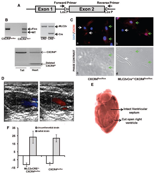
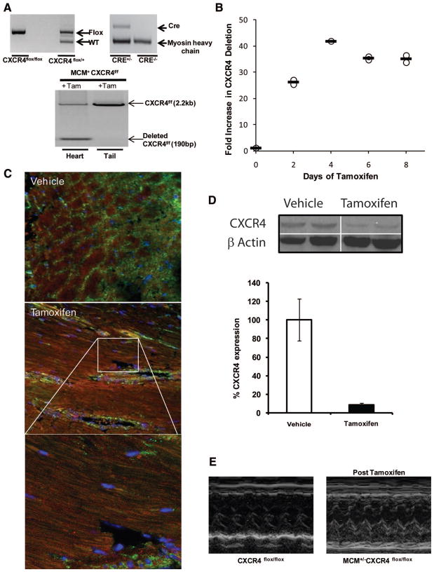
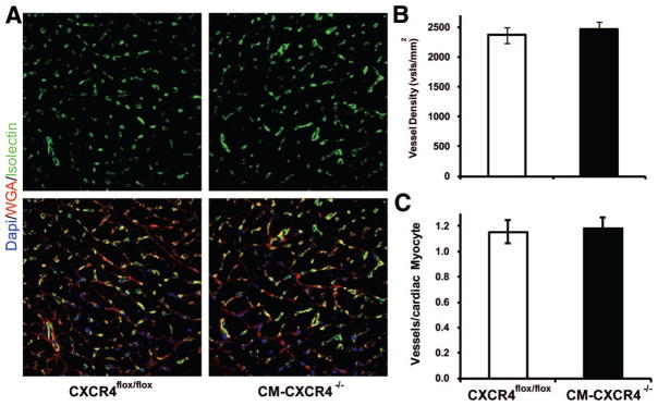
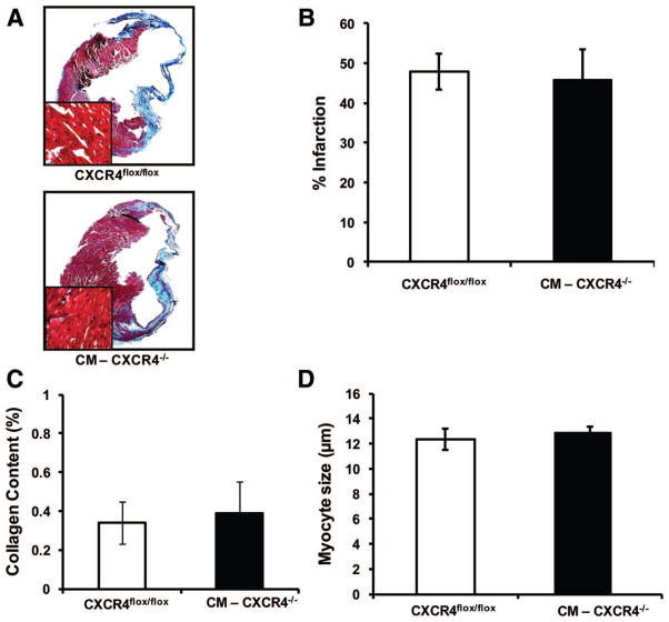
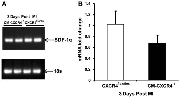
Methods and results: Two strains of CM-CXCR4(flox/flox) mice were generated by crossing CXCR4(flox/flox) mice with MCM-Cre(+/-) mouse and MLC2v-Cre(+/-) mouse on the C57BL/6J background, yielding CXCR4(flox/flox) MCM-Cre(+/-) and CXCR4(flox/flox)MLC2v-Cre(+/-) mice. Studies demonstrated recombination in both models congenitally in the MLC2v-Cre(+/-) mice and following tamoxifen administration in the MCM-Cre(+/-) mice. Surprisingly the CXCR4(flox/flox)MLC2v-Cre(+/-) are viable, had normal cardiac function, and had no evidence of ventricular septal defect. CXCR4(flox/flox)MCM(+/-) treated with tamoxifen 2 weeks before AMI demonstrated 90% decrease in cardiac CXCR4 expression 48 hours after AMI. Twenty-one days post AMI, echocardiography revealed no statistically significant difference in the wall thickness, left ventricular dimensions or ejection fraction (40.9+/-7.5 versus 34.4+/-2.6%) in CXCR4(flox/flox) mice versus CM-CXCR4(-/-) mice regardless of strategy of Cre expression. No differences in vascular density (2369+/-131 versus 2471+/-126 vessels/mm(2); CXCR4(flox/flox) versus CM-CXCR4(-/-) mouse), infarct size, collagen content, or noninfarct zone cardiac myocyte size were observed 21 days after AMI.
Conclusions: We conclude that cardiac myocyte-derived CXCR4 is not essential for cardiac development and, potentially because of the mismatch in timings of peaks of SDF-1 and CXCR4, has no major role in ventricular remodeling after AMI.
Figures

References
-
- Assmus B, Schachinger V, Teupe C, Britten M, Lehmann R, Dobert N, Grunwald F, Aicher A, Urbich C, Martin H, Hoelzer D, Dimmeler S, Zeiher AM. Transplantation of Progenitor Cells and Regeneration Enhancement in Acute Myocardial Infarction (TOPCARE-AMI) Circulation. 2002;106:3009–3017. - PubMed
-
- Britten MB, Abolmaali ND, Assmus B, Lehmann R, Honold J, Schmitt J, Vogl TJ, Martin H, Schachinger V, Dimmeler S, Zeiher AM. Infarct remodeling after intracoronary progenitor cell treatment in patients with acute myocardial infarction (TOPCARE-AMI): mechanistic insights from serial contrast-enhanced magnetic resonance imaging. Circulation. 2003;108:2212–2218. - PubMed
-
- Schachinger V, Erbs S, Elsasser A, Haberbosch W, Hambrecht R, Holschermann H, Yu J, Corti R, Mathey DG, Hamm CW, Suselbeck T, Assmus B, Tonn T, Dimmeler S, Zeiher AM. Intracoronary bone marrow-derived progenitor cells in acute myocardial infarction. N Engl J Med. 2006;355:1210–1221. - PubMed
-
- Brunner S, Winogradow J, Huber BC, Zaruba MM, Fischer R, David R, Assmann G, Herbach N, Wanke R, Mueller-Hoecker J, Franz WM. Erythropoietin administration after myocardial infarction in mice attenuates ischemic cardiomyopathy associated with enhanced homing of bone marrow-derived progenitor cells via the CXCR-4/SDF-1 axis. FASEB J. 2009;23:351–361. - PubMed
-
- Askari A, Unzek S, Popovic ZB, Goldman CK, Forudi F, Kiedrowski M, Rovner A, Ellis SG, Thomas JD, DiCorleto PE, Topol EJ, Penn MS. Effect of stromal-cell-derived factor-1 on stem cell homing and tissue regeneration in ischemic cardiomyopathy. Lancet. 2003;362:697–703. - PubMed
Publication types
MeSH terms
Substances
Grants and funding
LinkOut - more resources
Full Text Sources
Other Literature Sources
Medical
Molecular Biology Databases
