

Short communication: vascular smooth muscle cell stiffness as a mechanism for increased aortic stiffness with aging
- PMID: 20634486
- PMCID: PMC2936100
- DOI: 10.1161/CIRCRESAHA.110.221846
Short communication: vascular smooth muscle cell stiffness as a mechanism for increased aortic stiffness with aging
Abstract
Rationale: Increased aortic stiffness, an important feature of many vascular diseases, eg, aging, hypertension, atherosclerosis, and aortic aneurysms, is assumed because of changes in extracellular matrix (ECM).
Objective: We tested the hypothesis that the mechanisms also involve intrinsic stiffening of vascular smooth muscle cells (VSMCs).
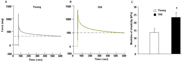
Methods and results: Stiffness was measured in vitro both by atomic force microscopy (AFM) and in a reconstituted tissue model, using VSMCs from aorta of young versus old male monkeys (Macaca fascicularis) (n=7/group), where aortic stiffness increases by 200% in vivo. The apparent elastic modulus was increased (P<0.05) in old (41.7+/-0.5 kPa) versus young (12.8+/-0.3 kPa) VSMCs but not after disassembly of the actin cytoskeleton with cytochalasin D. Stiffness of the VSMCs in the reconstituted tissue model was also higher (P<0.05) in old (23.3+/-3.0 kPa) than in young (13.7+/-2.4 kPa).
Conclusions: These data support the novel concept, not appreciated previously, that increased vascular stiffness with aging is attributable not only to changes in ECM but also to intrinsic changes in VSMCs.
Figures


References
-
- Mitchell GF, Guo CY, Benjamin EJ, Larson MG, Keyes MJ, Vita JA, Vasan RS, Levy D. Cross-sectional correlates of increased aortic stiffness in the community: the Framingham Heart Study. Circulation. 2007;20:2628–2636. - PubMed
-
- Qiu H, Depre C, Ghosh K, Resuello RG, Natividad FF, Rossi F, Peppas A, Shen YT, Vatner DE, Vatner SF. Mechanism of gender-specific differences in aortic stiffness with aging in nonhuman primates. Circulation. 2007;6:669–676. - PubMed
-
- Gaballa MA, Jacob CT, Raya TE, Liu J, Simon B, Goldman S. Large artery remodeling during aging: biaxial passive and active stiffness. Hypertension. 1998;3:437–443. - PubMed
-
- Karlon WJ, Hsu PP, Li S, Chien S, McCulloch AD, Omens JH. Measurement of orientation and distribution of cellular alignment and cytoskeletal organization. Ann Biomed Eng. 1999;6:712–720. - PubMed
Publication types
MeSH terms
Substances
Grants and funding
- R37 HL033107/HL/NHLBI NIH HHS/United States
- HL069752/HL/NHLBI NIH HHS/United States
- P01 AG027211/AG/NIA NIH HHS/United States
- HL069020/HL/NHLBI NIH HHS/United States
- R01 HL062863/HL/NHLBI NIH HHS/United States
- HL1024720/HL/NHLBI NIH HHS/United States
- AG027211/AG/NIA NIH HHS/United States
- AG023567/AG/NIA NIH HHS/United States
- P01 HL069020/HL/NHLBI NIH HHS/United States
- R01 AG023567/AG/NIA NIH HHS/United States
- R01 HL093481/HL/NHLBI NIH HHS/United States
- R01 HL102472/HL/NHLBI NIH HHS/United States
- R01 HL058690/HL/NHLBI NIH HHS/United States
- HL033107/HL/NHLBI NIH HHS/United States
- HL095888/HL/NHLBI NIH HHS/United States
- T32 HL069752/HL/NHLBI NIH HHS/United States
- R01 HL095888/HL/NHLBI NIH HHS/United States
- R01 HL033107/HL/NHLBI NIH HHS/United States
- JL 58960/PHS HHS/United States
LinkOut - more resources
Full Text Sources
Medical
Miscellaneous
