. 2010 Nov;95(11):1832-40.
doi: 10.3324/haematol.2010.027003. Epub 2010 Jul 15.

Transferrin receptor 2 and HFE regulate furin expression via mitogen-activated protein kinase/extracellular signal-regulated kinase (MAPK/Erk) signaling. Implications for transferrin-dependent hepcidin regulation

Affiliations

Transferrin receptor 2 and HFE regulate furin expression via mitogen-activated protein kinase/extracellular signal-regulated kinase (MAPK/Erk) signaling. Implications for transferrin-dependent hepcidin regulation

Maura Poli et al. Haematologica. 2010 Nov.

Abstract

Background: Impaired regulation of hepcidin in response to iron is the cause of genetic hemochromatosis associated with defects of HFE and transferrin receptor 2. However, the role of these proteins in the regulation of hepcidin expression is unclear.

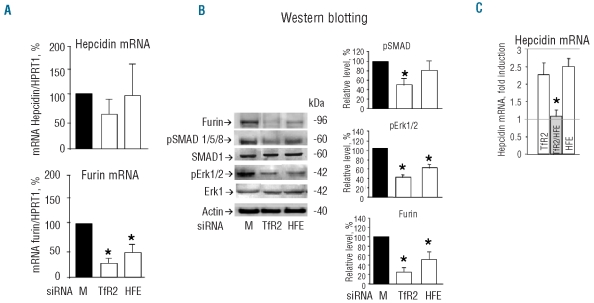
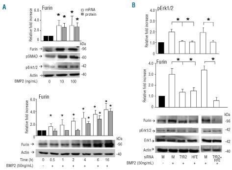
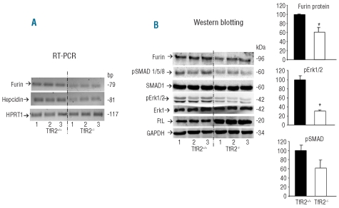
Design and methods: Hepcidin expression, SMAD and extracellular signal-regulated kinase (Erk) phosphorylation and furin expression were analyzed in hepatic HepG2 cells in which HFE and transferrin receptor 2 were down-regulated or expressed, or furin activity specifically inhibited. Furin expression was also analyzed in the liver of transferrin receptor 2 null mice.

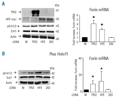
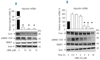
Results: We showed that the silencing of HFE and transferrin receptor 2 reduced both Erk phosphorylation and furin expression, that the exogenous expression of the two enhanced the induction of phosphoErk1/2 and furin by holotransferrin, but that this did not occur when the pathogenic HFE mutant C282Y was expressed. Furin, phosphoErk1/2 and phosphoSMAD1/5/8 were down-regulated also in transferrin receptor 2-null mice. Treatment of HepG2 cells with an inhibitor of furin activity caused a strong suppression of hepcidin mRNA, probably due to the inhibition of bone morphogenic protein maturation.

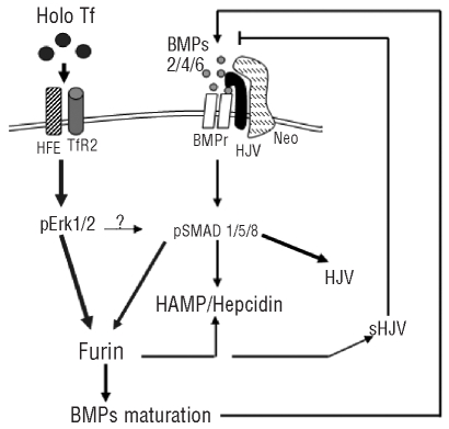
Conclusions: The data indicate that transferrin receptor 2 and HFE are involved in holotransferrin-dependent signaling for the regulation of furin which involved Erk phosphorylation. Furin in turn may control hepcidin expression.


Figures

Figure 1.
Figure 1.
Effects of TfR2 and HFE silencing. HepG2 cells were transfected with the siRNA specific for TfR2 and HFE and analyzed after 72 h. (A) Levels of hepcidin and furin mRNA evaluated by real-time RT-PCR after the transfection, expressed as percentages of the levels of the mock transfected cells corrected for HPRT1 mRNA level. (B) Western blotting of total cell homogenates probed with the antibodies for furin, phosphorylated SMAD1/5/8 (pSMAD1/5/8), SMAD1, phosphorylated Erk1/2 (pErk1/2), total Erk1 and actin; the histograms show the densitometry values of the bands expressed as percentages of those of the mock transfected cells, and corrected for actin level. (C) Real time analysis of hepcidin mRNA level after transfection with TfR2 or HFE siRNA alone and in combination, and after 50 ng/mL BMP2 for 16 h. The histogram is expressed as the percentage of hepcidin mRNA of the mock transfected cells. Histograms of the densitometry and of qRT-PCR are the means and SD of at least three independent experiments. The horizontal lines indicate the hepcidin mRMA level in the basal and in the induced control cells. The asterisks indicate statistically significant difference (P<0.05) from the mock transfected controls.
Figure 2.
Figure 2.
Treatment of HepG2 cells with BMP2. (A) Upper: HepG2 cells were treated with different doses of BMP2 (10–100 ng/mL) for 16 h and analyzed for furin mRNA with real-time RT-PCR and for furin, phosphoErk1/2, pSMAD1/5/8 and actin with western blotting. Lower: time course of furin induction by BMP2. HepG2 cells were grown in 50 ng/mL BMP2 and analyzed at the indicated times for furin mRNA with real-time RT-PCR and for furin and actin with western blotting. Histograms of the densitometry and of qRT-PCR are expressed as fold increase relative to the cells untreated with BMP2, after normalization on actin level, or HPRT1. The histograms are mean and SD of three independent experiments. The asterisks indicate statistically significant difference (P<0.05) from the mock transfected controls. (B) HepG2 cells were transfected with siRNA for TfR2 and HFE alone or in combination (TfR2+HFE), then they were incubated with 50 ng/mL BMP2 for 16 h, and furin and pErk1/2 were analyzed by western blotting. Histograms of the densitometry expressed as percentage relative to the mock transfected cells and incubated with BMP2 after normalization on actin level. Means and SD of at least three independent experiments. The asterisks indicate statistically significant difference (P<0.05) from the mock transfected controls.
Figure 3.
Figure 3.
Effect of holotransferrin in cells expressing HFE and TfR2. (A) The HepG2 cells were transfected with cDNA for TfR2 (TfR2), myc-tagged HFE (HFE) and myc-tagged HFE mutant C282Y (282). Right: western blotting analysis for the expression of the transgene with antibodies for TfR2 and Myc-Tag, pErk1/2 and total Erk1. Left: real time analysis of furin mRNA level after transfection with cDNA for TfR2 (TfR2), myc-tagged HFE and myc-tagged HFE mutant C282Y. (B) The transfected cells were incubated for 30 min with 30 μM holo-transferrin (HoloTf) and analyzed for pErk1/2 and total Erk1 level by western blotting (right). Left: real-time RT-PCR evaluation of furin mRNA after 30 min of incubation with holotransferrin. Western blots are representative of three independent experiments, and the histograms are means of three experiments. The asterisks indicate statistically significant difference (P<0.05) from the mock transfected controls (M).
Figure 4.
Figure 4.
Furin expression in TfR2−/− mice. The livers of three wild type (TfR2+/+) and of three TfR2 knockout (TfR2−/−) 14-day old mice were analyzed. (A) RT-PCR analysis of furin, hepcidin and HRPT1 (as a control) mRNA. (B) Western blotting analysis of the liver extracts for furin, pSMA1/5/8, total SMAD1, pErk1/2, total Erk1 and ferritin light chain (FtL). GAPDH was used as a loading control. The histograms represent the means of two groups analyzed, TfR2+/+ and TfR2−/−. The asterisks indicate a statistically significant difference (P<0.05).
Figure 5.
Figure 5.
Inhibition of furin activity. (A) HepG2 cells were incubated for 16 h with the indicated concentrations of furin inhibitor CMK and then the level of hepcidin mRNA analyzed by qRT-PCR, and pSMAD1/5/8, SMAD1, actin and furin analyzed by western blotting. (B) Cells were exposed to 50 μM CMK for the indicated time and the levels of hepcidin mRNA and of pSMAD1/5/8, SMAD1 furin and actin were analyzed. Histograms of qRT-PCR are the means and SD of at least three independent experiments. The asterisks indicate statistically significant difference (P<0.05) from the untreated cells (0).
Figure 6.
Figure 6.
Treatment with dorsomorphin and U0126. HepG2 cells were grown for 6 h in the presence or absence of 50 ng/mL BMP2 with or without 5 μM dorsomorphin (DM) or 10 μM U0126. (A) Western blot analysis of furin, pSMAD1/5/8, pErk1/2 and actin. Representative of three independent experiments. (B) Evaluation of hepcidin, and furin mRNA by real time RT-PCR. Histograms are expressed as fold-increase relative to non-treated cells. Means of three independent experiments.
Figure 7.
Figure 7.
Proposed scheme of the signaling pathway by TfR2 and HFE. Holotransferrin by binding to TfR2 in a complex with HFE, induces Erk1/2 phosphorylation. This, in turn, induces furin expression possibly acting also on the SMAD1/5/8 pathway. Furin participates in the maturation of hepcidin, and of the BMP, which induce hepcidin expression. It also produces the soluble form of HJV, which has an inhibitory effect on hepcidin expression.

References

    1. Ganz T. Hepcidin–a regulator of intestinal iron absorption and iron recycling by macrophages. Best Pract Res Clin Haematol. 2005;18(2):171–82. - PubMed
    1. Andrews NC. Forging a field: the golden age of iron biology. Blood. 2008;112(2):219–30. - PMC - PubMed
    1. Wang RH, Li C, Xu X, Zheng Y, Xiao C, Zerfas P, et al. A role of SMAD4 in iron metabolism through the positive regulation of hepcidin expression. Cell Metab. 2005;2(6):399–409. - PubMed
    1. Andriopoulos B, Jr, Corradini E, Xia Y, Faasse SA, Chen S, Grgurevic L, et al. BMP6 is a key endogenous regulator of hepcidin expression and iron metabolism. Nat Genet. 2009;41(4):482–7. - PMC - PubMed
    1. Zhang AS, West AP, Jr, Wyman AE, Bjorkman PJ, Enns CA. Interaction of hemojuvelin with neogenin results in iron accumulation in human embryonic kidney 293 cells. J Biol Chem. 2005;280(40):33885–94. - PubMed

Publication types

MeSH terms