. 2010 Aug;11(8):612-8.
doi: 10.1038/embor.2010.98. Epub 2010 Jul 16.

The structural plasticity of SCA7 domains defines their differential nucleosome-binding properties

Affiliations

The structural plasticity of SCA7 domains defines their differential nucleosome-binding properties

Jacques Bonnet et al. EMBO Rep. 2010 Aug.

Abstract

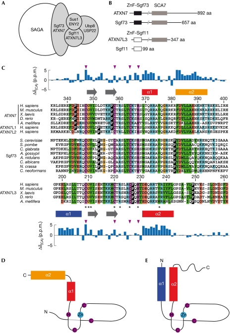
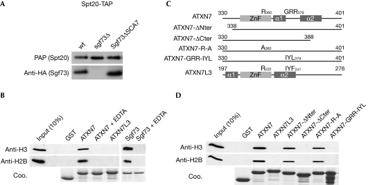
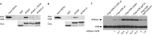
SAGA (Spt-Ada-Gcn5 acetyltransferase), a coactivator complex involved in chromatin remodelling, harbours both histone acetylation and deubiquitination activities. ATXN7/Sgf73 and ATXN7L3, two subunits of the SAGA deubiquitination module, contain an SCA7 domain characterized by an atypical zinc-finger. We show that the yeast Sgf73-SCA7 domain is not required to recruit Sgf73 into SAGA. Instead, it binds to nucleosomes, a property that is conserved in the human ATXN7-SCA7 domain but is lost in the ATXN7L3 domain. The solution structures of the SCA7 domain of both ATXN7 and ATXN7L3 reveal a new, common zinc-finger motif at the heart of two distinct folds, providing a molecular basis for the observed functional differences.


Conflict of interest statement

The authors declare that they have no conflict of interest.

Figures

Figure 1
Figure 1
Conservation of the SCA7 domain in ATXN7 and ATXN7L3 orthologues. (A) Schematic representation of the SAGA deubiquitination module. (B) Domain organization of ATXN7 homologues. The highly conserved SCA7 domain (grey box) is shared by all ATXN7 orthologues, as well as a subset of ATXN7L3 orthologues. Sequence conservation of the amino-terminal ZnF domain distinguishes ATXN7L3/Sgf11 orthologues (ZnF-Sgf11, white box) from ATXN7/Sgf73 orthologues (ZnF-Sgf73, black box). (C) Multiple sequence alignment of the SCA7 domain from selected species reveals the sequence similarities and differences that allow the SCA7 domains of ATXN7/Sgf73 orthologues to be distinguished from those of ATXN7L3 orthologues. Invariant residues are indicated by asterisks and zinc-chelating residues are coloured in magenta. Hydrophobic, positive, negative and glycine residues are coloured in green, blue, red and yellow, respectively. Prolines are shown in inverted contrast. α-Helices are indicated as rectangles and extended segments as arrows, and the deviations of Cα chemical shifts from random coil values are represented above and below the ATXN7 and ATXN7L3 sequences. The positions of zinc-coordinating residues are indicated with magenta triangles. (D,E) Topologies of ATXN7–SCA7 (D) and ATXN7L3–SCA7 domains (E). SAGA, Spt–Ada–Gcn5 acetyltransferase; ZnF, zinc-finger.
Figure 2
Figure 2
Solution structures of the SCA7 domains of human ATXN7 and ATXN7L3. (A,B) Ribbon representation of (A) ATXN7–SCA7 and (B) ATXN7L3–SCA7 domains showing the hydrophobic contacts that hook the two α-helices onto the zinc-finger. (C,D) Conserved interactions define the relative orientations of the two α-helices within (C) ATXN7–SCA7 and (D) ATXN7L3–SCA7 domains. The side chains of conserved hydrophobic, positive or negative residues are shown by using green, blue or red sticks, respectively. The figures were prepared using PyMOL (DeLano Scientific LLC).
Figure 3
Figure 3
The SCA7 domains of human ATXN7 and yeast Sgf73 bind to nucleosomes. (A) Tandem affinity purifications (TAP) of SAGA using TAP-tagged Spt20 from the indicated strains were analysed by western blot using PAP or HA antibody. (B) SCA7 domains from ATXN7, ATXN7L3 or Sgf73 fused to GST or GST alone were immobilized on glutathione-Sepharose beads and incubated with mononucleosomes. The retained material was analysed by Coomassie blue staining (Coo.) and western blotting using H2B or H3 antibodies, as indicated. All interactions were lost when the fusion proteins were pre-incubated with EDTA solution. (C) Schematic representation of ATXN7–SCA7 mutant domains used in GST-pulldown experiments. (D) Mononucleosomes retained by immobilized SCA7 domains were analysed by western blotting using H3 and H2B antibodies. GST, glutathione S-transferase; HA, haemagglutinin; PAP, peroxidase anti-peroxidase; SAGA, Spt–Ada–Gcn5 acetyltransferase; ZnF, zinc-finger.
Figure 4
Figure 4
The yeast Sgf73–SCA7 domain regulates H2B deubiquitination. (A,B) GST-pulldown experiments using H2A–H2B dimers or tail-less histone octamers were analysed by Coomassie blue staining (Coo.) or western blotting using H2B antibodies. (C) Extracts of the indicated yeast strains carrying a Flag–HTB1 plasmid were separated on a 15% SDS–PAGE gel and subjected to western blot analysis using Flag antibodies. GST, glutathione S-transferase; SDS–PAGE, SDS–polyacrylamide gel electrophoresis.

References

    1. Baker SP, Grant PA (2007) The SAGA continues: expanding the cellular role of a transcriptional co-activator complex. Oncogene 26: 5329–5340 - PMC - PubMed
    1. Bonnet J, Romier C, Tora L, Devys D (2008) Zinc-finger UBPs: regulators of deubiquitylation. Trends Biochem Sci 33: 369–375 - PubMed
    1. Cornilescu G, Delaglio F, Bax A (1999) Protein backbone angle restraints from searching a database for chemical shift and sequence homology. J Biomol NMR 13: 289–302 - PubMed
    1. Daniel JA, Torok MS, Sun ZW, Schieltz D, Allis CD, Yates JR III, Grant PA (2004) Deubiquitination of histone H2B by a yeast acetyltransferase complex regulates transcription. J Biol Chem 279: 1867–1871 - PubMed
    1. Helmlinger D et al. (2004) Ataxin-7 is a subunit of GCN5 histone acetyltransferase-containing complexes. Hum Mol Genet 13: 1257–1265 - PubMed

Publication types

MeSH terms