Review
. 2008;1(2008):23-40.
doi: 10.1109/RBME.2008.2008233. Epub 2008 Nov 5.

Multimodal functional neuroimaging: integrating functional MRI and EEG/MEG

Affiliations
Review

Multimodal functional neuroimaging: integrating functional MRI and EEG/MEG

Bin He et al. IEEE Rev Biomed Eng. 2008.

Abstract

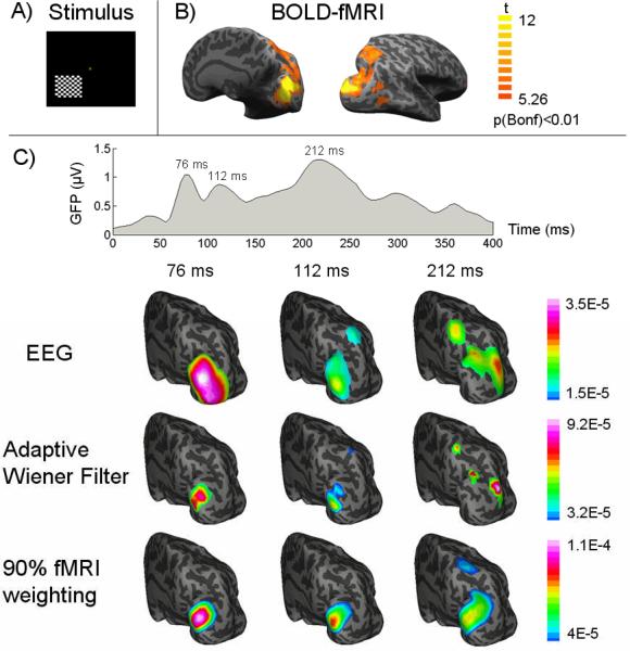
Noninvasive functional neuroimaging, as an important tool for basic neuroscience research and clinical diagnosis, continues to face the need of improving the spatial and temporal resolution. While existing neuroimaging modalities might approach their limits in imaging capability mostly due to fundamental as well as technical reasons, it becomes increasingly attractive to integrate multiple complementary modalities in an attempt to significantly enhance the spatiotemporal resolution that cannot be achieved by any modality individually. Electrophysiological and hemodynamic/metabolic signals reflect distinct but closely coupled aspects of the underlying neural activity. Combining fMRI and EEG/MEG data allows us to study brain function from different perspectives. In this review, we start with an overview of the physiological origins of EEG/MEG and fMRI, as well as their fundamental biophysics and imaging principles, we proceed with a review of the major advances in the understanding and modeling of neurovascular coupling and in the methodologies for the fMRI-EEG/MEG simultaneous recording. Finally, we summarize important remaining issues and perspectives concerning multimodal functional neuroimaging, including brain connectivity imaging.

Keywords: EEG; MEG; Multimodal neuroimaging; fMRI; human brain mapping; neurovascular coupling.


Figures

Figure 1
Figure 1
Schematic illustration of the ranges of spatial and temporal resolution of various noninvasive (in blue) imaging techniques and invasive (in red) experimental techniques.
Figure 2
Figure 2
Illustration of the fMRI-EEG/MEG integrated multimodal neuroimaging (part of figure adapted from Fig. 1 of [46] with permission).
Figure 3
Figure 3
Schematical illustration of BOLD-contrast fMRI. The regional neuronal activity alters the local CBF, CBV and CMRO2, which collectively leads to changes in the blood oxygen level. The increase of oxygen level, meaning the decrease of local field inhomogeniety, produces a longer T2 or T2* and therefore larger MR signals. The frequency-and-phase encoding technique (e.g. echo-planar imaging) allows for the fast acquisition of a so-called k-space data, which can be transformed to the original image space through Fourier transformation. The signal is most frequently analyzed voxel by voxel, yielding statistic maps indicating regions with significant hemodynamic effects related to external stimuli/tasks or internal events. These regions arguably define the activated neuronal populations.
Figure 4
Figure 4
The BOLD fMRI signal at an “activated” voxel can be modeled using a linear system. The signal is the scaled version of a location-independent predictor signal, derived from the stimulus function and the hemodynamic response function (HRF). The scaling factor (called the BOLD effect size) is proportional to the time integral of the event-related synaptic power.
Figure 5
Figure 5
Flow-chart of the fMRI-EEG/MEG co-registration.
Figure 6
Figure 6
A) The pattern-reversal checkerboard visual stimulation, B) fMRI activation map with a corrected threshold p<0.01, and C) the global field power of VEP and the dynamic cortical source distribution at three VEP latencies (76, 112, 212 ms after the visual onset) imaged from EEG alone (1st row), or fMRI-EEG integration using our proposed adaptive wiener filter (2nd row) and the conventional 90% fMRI weighted algorithm (3rd row). Both the source images and the fMRI activation map are visualized on an inflated representation of cortical surface. (From [24] with permission)
Figure 7
Figure 7
Typical experimental setting for simultaneous fMRI-EEG recordings.
Figure 8
Figure 8
Examples of simultaneous fMRI-EEG data in three control experiments. A) VEP waveforms obtained inside the fMRI scanner with (blue) or without (dashed red) fMRI scans, or outside the scanner (black). The scalp potential map corresponds to the 120-ms peak latency in the VEP acquired during fMRI scans. B) SSVEP power spectrum and the spatial distribution of the power at the stimulus frequency. C) Time-frequency representation of the alpha modulation induced by self-paced eye-open and eye-close. (Panel A) from [174] with permission)

References

    1. Berger H. Ü ber das Elektroenkephalogram des Menschen. Arch. f. Psychiat. 1929;87:527–70.
    1. Cohen D. Magnetoencephalography: evidence of magnetic fields produced by alpha-rhythm currents. Science. 1968 Aug 23;161:784–786. - PubMed
    1. Ogawa S, Tank DW, Menon R, Ellermann JM, Kim SG, Merkle H, Ugurbil K. Intrinsic signal changes accompanying sensory stimulation: functional brain mapping with magnetic resonance imaging. Proc. Natl. Acad. Sci. U. S. A. 1992 Jul 1;89:5951–5955. - PMC - PubMed
    1. Kwong KK, Belliveau JW, Chesler DA, Goldberg IE, Weisskoff RM, Poncelet BP, Kennedy DN, Hoppel BE, Cohen MS, Turner R. Dynamic magnetic resonance imaging of human brain activity during primary sensory stimulation. Proc. Natl. Acad. Sci. U. S. A. 1992 Jun 15;89:5675–5679. - PMC - PubMed
    1. Bandettini PA, Wong EC, Hinks RS, Tikofsky RS, Hyde JS. Time course EPI of human brain function during task activation. Magn. Reson. Med. 1992 Jun;25:390–397. - PubMed

Publication types

MeSH terms