. 2010 Jul 12;5(7):e11524.
doi: 10.1371/journal.pone.0011524.

Visualizing early splenic memory CD8+ T cells reactivation against intracellular bacteria in the mouse

Affiliations

Visualizing early splenic memory CD8+ T cells reactivation against intracellular bacteria in the mouse

Marc Bajénoff et al. PLoS One. .

Abstract

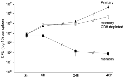
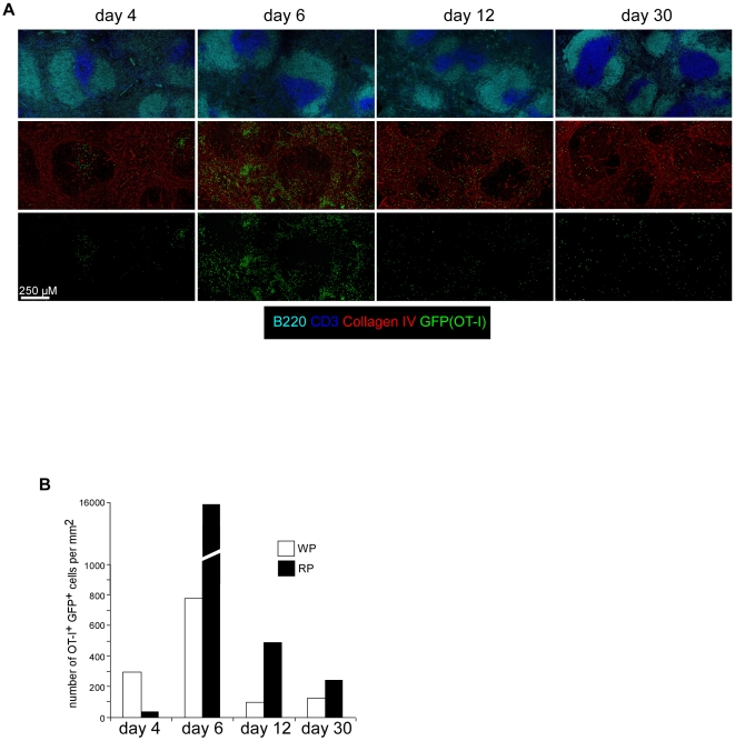
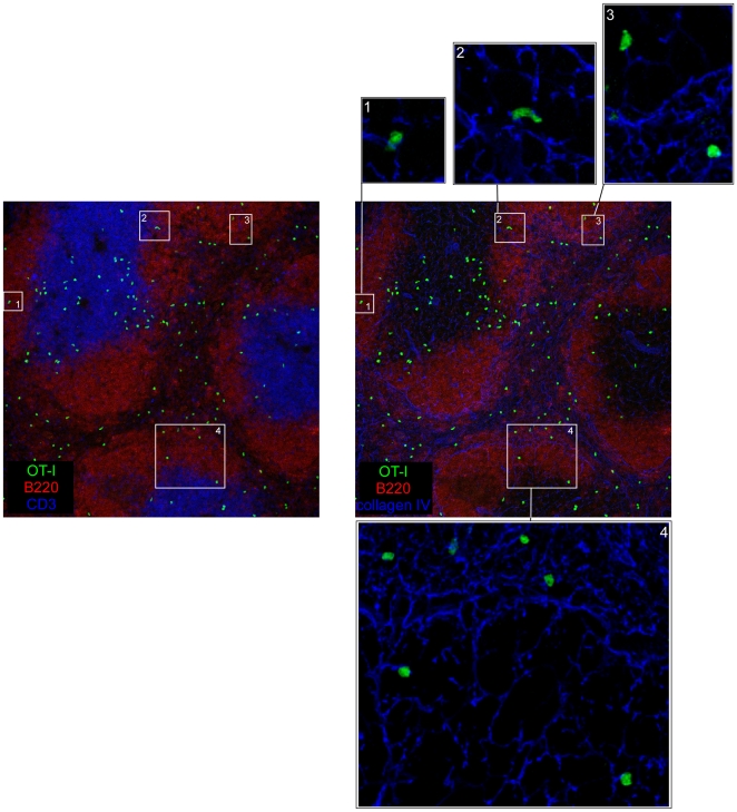
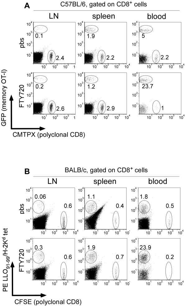
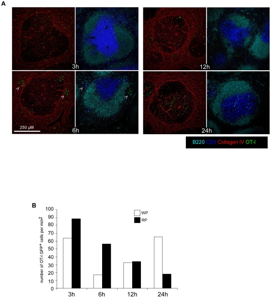
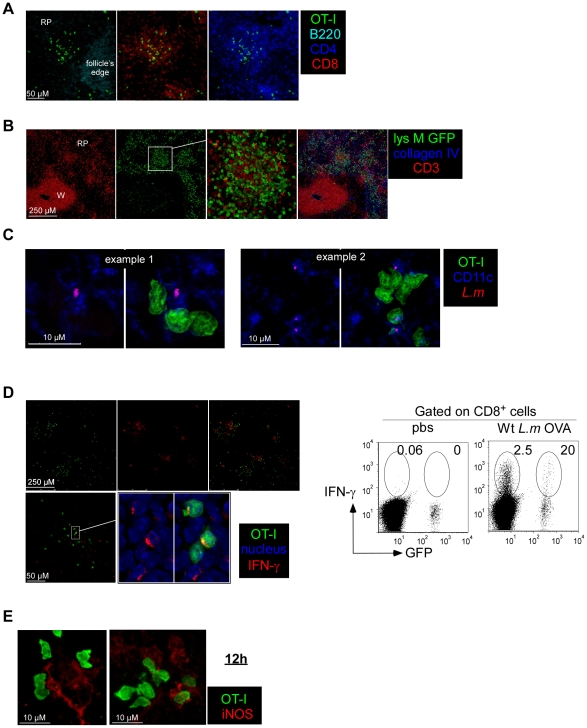
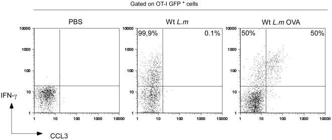
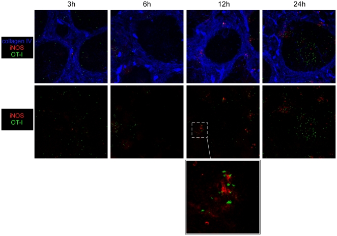
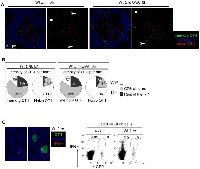
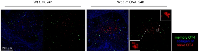
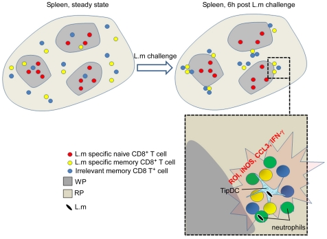
Memory CD8(+) T cells represent an important effector arm of the immune response in maintaining long-lived protective immunity against viruses and some intracellular bacteria such as Listeria monocytogenes (L.m). Memory CD8(+) T cells are endowed with enhanced antimicrobial effector functions that perfectly tail them to rapidly eradicate invading pathogens. It is largely accepted that these functions are sufficient to explain how memory CD8(+) T cells can mediate rapid protection. However, it is important to point out that such improved functional features would be useless if memory cells were unable to rapidly find the pathogen loaded/infected cells within the infected organ. Growing evidences suggest that the anatomy of secondary lymphoid organs (SLOs) fosters the cellular interactions required to initiate naive adaptive immune responses. However, very little is known on how the SLOs structures regulate memory immune responses. Using Listeria monocytogenes (L.m) as a murine infection model and imaging techniques, we have investigated if and how the architecture of the spleen plays a role in the reactivation of memory CD8(+) T cells and the subsequent control of L.m growth. We observed that in the mouse, memory CD8(+) T cells start to control L.m burden 6 hours after the challenge infection. At this very early time point, L.m-specific and non-specific memory CD8(+) T cells localize in the splenic red pulp and form clusters around L.m infected cells while naïve CD8(+) T cells remain in the white pulp. Within these clusters that only last few hours, memory CD8(+) T produce inflammatory cytokines such as IFN-gamma and CCL3 nearby infected myeloid cells known to be crucial for L.m killing. Altogether, we describe how memory CD8(+) T cells trafficking properties and the splenic micro-anatomy conjugate to create a spatio-temporal window during which memory CD8(+) T cells provide a local response by secreting effector molecules around infected cells.


Conflict of interest statement

Competing Interests: The authors have declared that no competing interests exist.

Figures

Figure 1
Figure 1. Memory CD8 T cells rapidly control secondary L.m infection.
Mice (3 per group) were infected i.v with PBS or immunized with 0.1×LD50 Wt L.m bacteria. 30 days later, animals were injected or not with 100 µg of anti-CD8β depleting Ab i.p for three consecutive days and then challenged with 7×10⁵ Wt bacteria. Bacteria titers in the spleen were measured 3, 6, 24 and 48 hours later. Data are representative of 2 independent experiments.
Figure 2
Figure 2. Within the spleen, L.m specific memory CD8 T+ cells predominantly reside in the Red Pulp.
(A, B) 200 naïve GFP+ OT-I cells were transferred in C57BL/6 mice. One day after, recipient mice were injected i.v. with 0.1×LD50 Wt L.m-OVA. Spleens were harvested 4, 6, 12 and 30 days later, sectioned, stained for B220, CD3, collagen IV expression and analyzed by confocal microscopy. (B) The surfaces of WP and RP zones were delineated based on collagen IV expression and summed up. The densities of memory GFP+ OT-I cells per mm2 of each region were calculated and displayed. Data are representative of 3 independent experiments.
Figure 3
Figure 3. L.m-specific memory CD8+ T cells present in follicles are adjacent to reticular fibers.
200 naïve GFP+ OT-I cells were transferred in C57BL/6 mice that were injected i.v. the following day with 0.1×LD50 Wt L.m-OVA. Spleens were harvested 30 days later, sectioned, stained for B220, CD3, collagen IV expression and analyzed by confocal microscopy. Inserts show enlargements of B220+ areas containing OT-I memory cells. Data are representative of 2 different experiments.
Figure 4
Figure 4. L.m specific memory CD8+ T cells preferentially traffic in the bloodstream.
(A) 200 naïve GFP+ OT-I cells were transferred in C57BL/6 mice. One day later, recipient mice were injected i.v. with 0.1×LD50 Wt L.m-OVA. Thirty days after this immunization, recipient mice were injected i.v with 3×10⁶ naïve CMTPX-labelled polyclonal CD8+ T cells. The following day, mice were injected i.p with PBS or 100 µg of FTY720. Animals were killed 24 hours later and their LNs, spleen and blood harvested, stained for CD8 expression and analyzed by flow cytometry. Numbers indicate the percentages of GFP+ OT-I memory cells and CMTPX naive polyclonal CD8+ T cells among total CD8+ T cells. (B) Wt BALB/c mice were injected i.v with 0.1×LD50 Wt L.m and further treated as in (A) with the exception that polyclonal naive CD8+ T cells were labelled with CFSE and that L.m specific endogenous memory CD8+ T cells were identified as LLO9199/H-2Kd tetramers+ CD8+ cells. Data are representative of 3 independent experiments.
Figure 5
Figure 5. L.m-specific memory CD8+ T cells transiently form clusters in the Red Pulp of secondary infected animals.
(A, B) 200 naïve GFP+ OT-I cells were transferred in C57BL/6 mice that were injected i.v. the following day with 0.1×LD50 Wt L.m-OVA. One month later, mice were injected i.v. with PBS or 10×LD50 Wt L.m-OVA. Spleens were harvested 3, 6, 12 and 24 hours later, sectioned, stained for B220, CD3, collagen IV expression and analyzed by confocal microscopy. Arrowheads indicate clusters of GFP+ OT-I memory cells 6 hours post re-infection. (B) The surfaces of WP and RP zones were delineated based on collagen IV expression and summed up. The densities of memory GFP+ OT-I cells per mm2 of each region were calculated and displayed. Data are representative of 3 independent experiments.
Figure 6
Figure 6. Memory ‘CD8 clusters’ concentrate anti-listeria effector cells.
(A,C,D,E) 200 naïve GFP+ OT-I cells were transferred in C57BL/6 mice. One day after, recipient mice as well as naive GFP+ lys-M Tg mice (B) were injected i.v. with 0.1×LD50 Wt L.m-OVA. One month later, all mice were injected i.v. with 10×LD50 Wt L.m-OVA. Spleens were harvested 6 (A,B,C,D) or 12 hrs later (E), sectioned, stained with anti-B220, -CD4, -CD8 (A); anti-CD3, -collagen IV (B) anti-CD11c, -listeria (C); anti-IFN-γ, and hoescht (D), anti-iNOS (E) specific Abs and analyzed by confocal microscopy. In (D), IFN-γ secretion by endogenous CD8+ T cells and GFP+ OT-I memory cells was also assessed by flow cytometry. Data are representative of 3 independent experiments.
Figure 7
Figure 7. Both L.m-specific and non-specific memory CD8+ T cells produce IFN-γ, but only TCR-triggered cells secrete CCL3.
200 naïve GFP+ OT-I cells were transferred in C57BL/6 mice that were injected i.v. the following day with 0.1×LD50 Wt L.m-OVA. Thirty days after, recipient mice were injected with 10×LD50 Wt L.m or Wt L.m-OVA. Spleens were harvested 6 hours later, directly stained for IFN-γ and CCL3 expression and analyzed by flow cytometry. The numbers in the quadrants indicate the percentages of IFN-γ + GFP+ OT-I co-secreting or not CCL3.
Figure 8
Figure 8. Inflammatory monocytes are activated/recruited to the effector clusters.
200 naïve GFP+ OT-I cells were transferred in C57BL/6 mice that were infected i.v. the following day with 0.1×LD50 Wt L.m-OVA. Thirty days after, recipient mice were injected with 10×LD50 Wt L.m-OVA. Spleens were harvested 3, 6, 12 and 24 hours later, sectioned, stained for iNOS, collagen IV expression and analyzed by confocal microscopy. Insert shows an enlargement of an effector cluster. Data are representative of 2 different experiments.
Figure 9
Figure 9. L.m-specific and non-specific memory CD8+ T cells aggregate in effector clusters and secrete IFN-γ upon L.m reinfection.
200 naïve GFP+ OT-I cells were transferred in C57BL/6 mice that were injected i.v. the following day with 0.1×LD50 Wt L.m-OVA.. Thirty days after, recipient mice were injected with 3×10⁶ naïve CMTPX labelled OT-I cells. The following day, mice were injected i.v. with 10×LD50 Wt L.m or Wt L.m-OVA. Spleens were harvested 6 hours later, sectioned, stained for collagen IV (A) or IFN-γ (C) expression and analyzed by confocal microscopy. In C, IFN-γ secretion by endogenous CD8+ T cells and memory GFP+ OT-I cells was also assessed by flow cytometry. (B) Spleen sections were stained for collagen IV and CD8 expression. The surfaces of 3 regions of interest were drawn: the WP, the RP area containing endogenous CD8 clusters and the rest of the RP. The densities of memory GFP+ OT-I and naïve CMTPX OT-I cells per mm2 of each region of interest were calculated and displayed. Data are representative of 2 different experiments.
Figure 10
Figure 10. L.m-specific naïve CD8 T+ cells are reactivated in the WP.
200 naïve GFP+ OT-I cells were transferred in C57BL/6 mice that were injected i.v. the following day with 0.1×LD50 Wt L.m-OVA. One month later, recipient mice were injected i.v. with 3×10⁶ naïve CMTPX labelled OT-I cells. One day after, mice were injected i.v. with 10×LD50 Wt L.m or Wt L.m-OVA. Spleens were harvested 24 hrs later, sectioned, stained for collagen IV expression and analyzed by confocal microscopy. Inserts show the clusterization of naïve OT-I cells in the WP of Wt L.m-OVA challenged animals. Data are representative of 2 different experiments.
Figure 11
Figure 11. Summary.
At the steady state, while splenic L.m-specific naive CD8+ T cells reside in the WP, L.m specific memory cells preferentially traffic in the RP as a result of their increased patrolling of the blood system. Within six hours post L.m re-infection, clusters of cells rapidly form around L.m-infected cells in the splenic RP of challenged animals. These clusters contain several immune effector cells that express antimicrobial activities such as IFN-γ and CCL3 secreting memory CD8+ T cells, neutrophils and Tip-DCs. Importantly, the capacity to join these clusters and secrete IFN-γ is independent of TCR recognition but requires the CD8+ T cells to belong to the memory lineage. Because these clusters that involve many antimicrobial effector cells and molecules occur at the exact same time when L.m burden starts to be controlled by memory CD8+ T cells, we believe that they may be the first site where L.m infection is controlled during a recall splenic infection.

References

    1. Harty JT, Tvinnereim AR, White DW. CD8+ T cell effector mechanisms in resistance to infection. Annu Rev Immunol. 2000;18:275–308. - PubMed
    1. Willard-Mack CL. Normal structure, function, and histology of lymph nodes. Toxicol Pathol. 2006;34:409–424. - PubMed
    1. Young AJ. The physiology of lymphocyte migration through the single lymph node in vivo. Semin Immunol. 1999;11:73–83. - PubMed
    1. Bajenoff M, Egen JG, Qi H, Huang AY, Castellino F, et al. Highways, byways and breadcrumbs: directing lymphocyte traffic in the lymph node. Trends Immunol. 2007;28:346–352. - PubMed
    1. Bajenoff M, Granjeaud S, Guerder S. The strategy of T cell antigen-presenting cell encounter in antigen-draining lymph nodes revealed by imaging of initial T cell activation. J Exp Med. 2003;198:715–724. - PMC - PubMed

Publication types