. 2010 Sep;77(5):1123-35.
doi: 10.1111/j.1365-2958.2010.07276.x.

Molecular analysis of the prokaryotic ubiquitin-like protein (Pup) conjugation pathway in Mycobacterium tuberculosis

Affiliations

Molecular analysis of the prokaryotic ubiquitin-like protein (Pup) conjugation pathway in Mycobacterium tuberculosis

Francisca A Cerda-Maira et al. Mol Microbiol. 2010 Sep.

Abstract

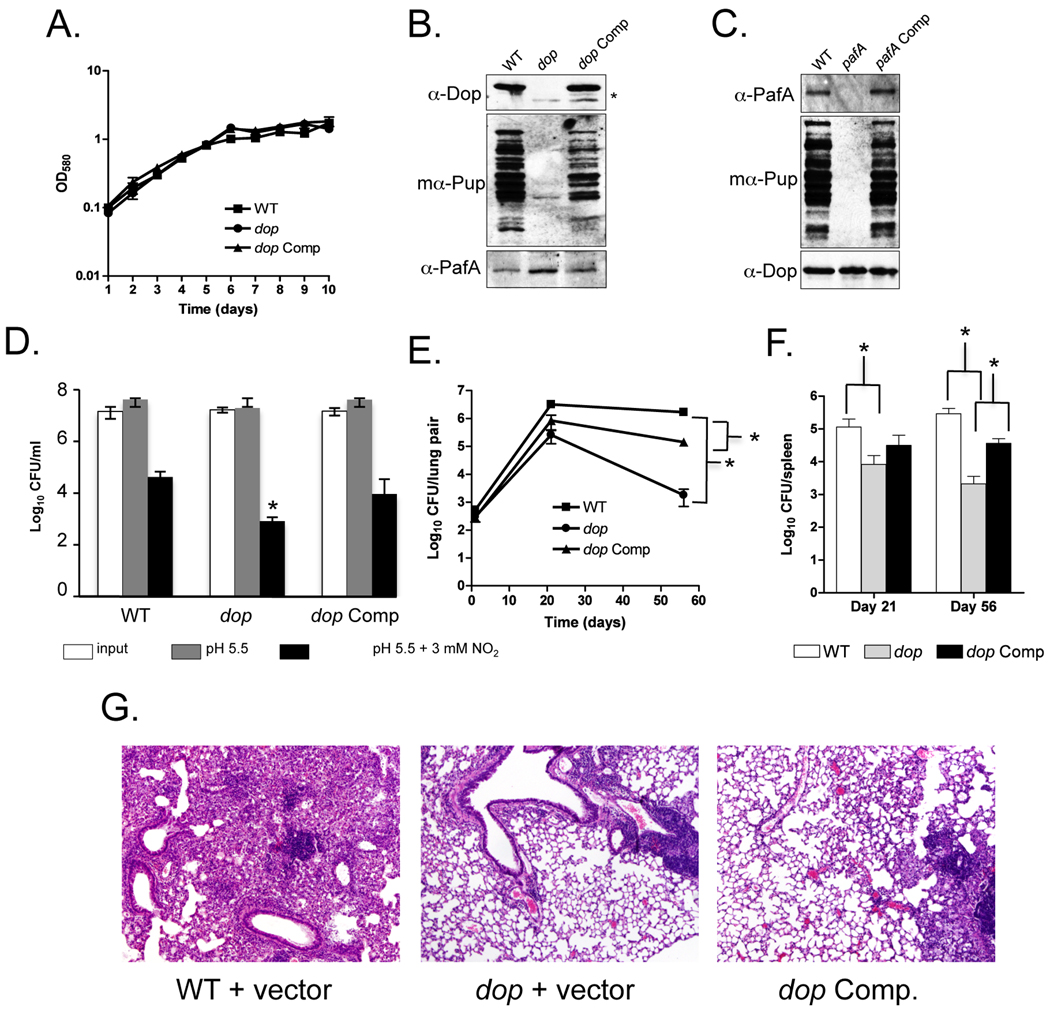
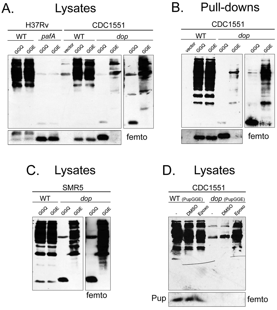
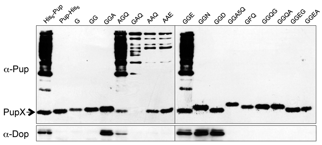
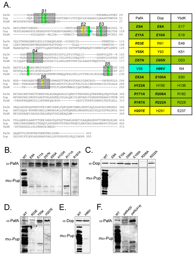
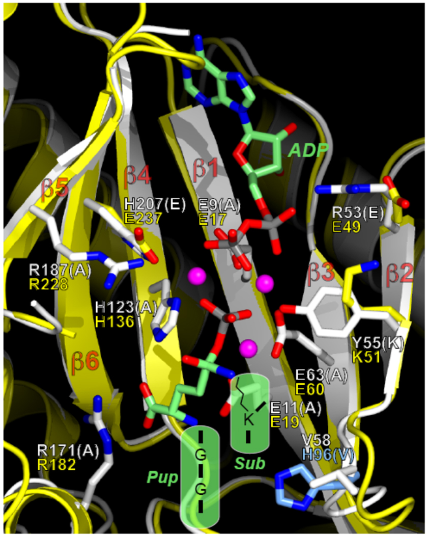
Proteins targeted for degradation by the Mycobacterium proteasome are post-translationally tagged with prokaryotic ubiquitin-like protein (Pup), an intrinsically disordered protein of 64 residues. In a process termed 'pupylation', Pup is synthesized with a terminal glutamine, which is deamidated to glutamate by Dop (deamidase of Pup) prior to attachment to substrate lysines by proteasome accessory factor A (PafA). Importantly, PafA was previously shown to be essential to cause lethal infections by Mycobacterium tuberculosis (Mtb) in mice. In this study we show that Dop, like PafA, is required for the full virulence of Mtb. Additionally, we show that Dop is not only involved in the deamidation of Pup, but also needed to maintain wild-type steady state levels of pupylated proteins in Mtb. Finally, using structural models and site-directed mutagenesis our data suggest that Dop and PafA are members of the glutamine synthetase fold family of proteins.


Figures

Fig. 1
Fig. 1
Mtb Dop is required for pupylation, resistance to RNI and virulence in mice. (A) Growth curve analysis of WT, dop and dop-complemented (“dop Comp”) Mtb CDC1551 strains. The strains were grown with no aeration and the optical density at absorbance 580 nm (OD580) of the cultures was measured daily. (B) An Mtb dop mutant is defective for pupylation. Total cell lysates from equivalent cell numbers of WT, dop, and dop-complemented ("dop Comp") CDC1551 strains were analyzed by immunoblotting. Asterisk (*) indicates endogenous truncated Dop resulting from the transposon insertion in dop. "mα-Pup" indicates the use of monoclonal antibodies to Pup. (C) Total cell lysates from equivalent cell numbers of WT, pafA, pafA-complemented ("pafA Comp") H37Rv strains. For (B) and (C), proteins were separated by 10% SDS-PAGE and analyzed with polyclonal antibodies to Dop or PafA, or monoclonal antibodies to Pup. (D) The Mtb dop mutant is hypersusceptible to RNI. Survival of WT, dop and dop-complemented Mtb CDC1551 strains was determined after six days of incubation in media pH 5.5 in the presence or absence of 3 mM NaNO2. Error bars indicate ± one standard deviation (SD). Asterisk (*) indicates the dop mutant was statistically significantly more sensitive to RNI than WT or the complemented strains (P < 0.05). Data are from two independent experiments each performed in triplicate, with an outlier excluded from each strain tested. (E, F) The dop Mtb mutant is severely attenuated for growth and survival in mice. Colony forming units (CFU) from lungs (E) and spleens (F) harvested on day 1 (n = 3), day 21 (n = 4) and day 56 (n = 4) from mice infected with WT, dop and dop-complemented Mtb CDC1551 strains. Error bars indicate ± one SD. Single asterisk (*) indicates a P value of < 0.05. The WT and dop-complemented strains were significantly different from each other in the lungs (as indicated) and spleens (not indicated due to space constraints), suggesting complementation was not complete. All statistics were performed using a nonparametric Student’s two-tailed t test. (G) Hematoxylin and eosin staining of formalin fixed lungs from mice infected with WT (MHD583), dop (MHD375) and dop-complemented (MHD376) Mtb CDC1551 strains. 4 5 magnification is shown.
Fig. 2
Fig. 2
Expression of pupGGE is not sufficient to bypass the requirement for Dop in Mtb. (A) Mtb H37Rv WT and pafA or CDC1551 WT and dop strains were transformed with plasmids encoding N-terminal His6 tagged pupGGQ, pupGGE or empty vector. Lysates with equivalent cell numbers from these strains were analyzed by 12% SDS-PAGE and immunoblotting with polyclonal antibodies to Pup. At least two transformants from each electroporation were characterized with similar results. (B) His6-PupGGQ or His6-PupGGE pupylomes from the same strains in (A) were purified. Proteins were analyzed by 12% SDS-PAGE and immunoblotting. (C) The same plasmids encoding pupGGQ and pupGGE were used to transform the previously reported Msm dop mutant and the parental WT strain. Total cell lysates with equivalent cell numbers were separated by 12% SDS-PAGE and analyzed with polyclonal antibodies to Pup. ThermoScientific Super Signal West Femto reagent (“femto”) was used to detect free Pup (lower panels) or the pupylomes in the dop mutant (right panels) for (A)–(C). (D) Proteasomal inhibition appeared to rescue pupylated proteins from degradation in the Mtb dop mutant expressing pupGGE.
Fig. 3
Fig. 3
Mutation analysis of the C-terminus of Pup. WT Msm was transformed with plasmids expressing his6-pup with different C-terminal mutations. His6-Pup C-terminus mutant proteins were purified from soluble lysates and analyzed by 12% SDS-PAGE gel and immunoblotting with polyclonal antibodies to Pup. Only the terminal residues are indicated for the mutant proteins. “PupX” indicates unconjugated WT or C-terminal mutant Pup. Immunoblotting with antibodies to Dop reveals Dop requires the di-glycine motif for robust binding to Pup (lower panel).
Fig. 4
Fig. 4
Dop and PafA share critical catalytic residues with GS-fold family members. (A) (Left) Alignment of PafA, Dop and E. coli YbdK shows conserved residues crucial for GS-fold protein function in β strands (boxed in grey). Highlighted residues were selected for mutagenesis. Sequences were compiled from the NCBI server and aligned in segments using ClustalW (http://www.ebi.ac.uk/Tools/clustalw2/index.html). (Right) Residues conserved among PafA, Dop and GS-fold protein members are highlighted in green. Amino acids conserved among PafA and Dop homologues, but not in other GS-fold members are highlighted in yellow. A residue conserved in Dop homologues but not in PafA homologues is in cyan. (B)–(C) Residues conserved in GS/γ-GCS-fold proteins are important for PafA (B) and Dop (C) function. Site-directed mutation analysis of PafA and Dop. Null mutants of pafA or dop were transformed by electroporation with plasmids encoding pafA or dop, respectively, with single mutations. In some samples, a cross-reactive band is seen in the pafA mutant lysates below PafA. (D) Residues conserved in PafA and Dop homologues but not found in GS-fold proteins are critical for PafA function. (E) Replacement of a conserved histidine in Dop homologues for a valine conserved in PafA family members eliminates pupylation. (F) The C-terminus of PafA is required for pupylation. Endogenous PafA420 and recombinant PafA414 are indicated. For all experiments, total cell lysates of equivalent cell numbers were separated by 10% SDS-PAGE and analyzed by immunoblot analysis with antibodies to PafA, Dop or Pup as in Fig. 1.
Fig. 5
Fig. 5
Proposed model of the PafA/Dop active site. Superimposition of the structural model of PafA with the template structure of E. coli YbdK (PDB code 1R8G) (Lehmann et al., 2004). Large insertions or deletions in PafA relative to YbdK were truncated due to the model’s poor reliability in these regions. The secondary-structure elements and side-chain carbon atoms of PafA are coloured white, and those for YbdK are coloured yellow. Select side chains for PafA and YbdK are shown in stick representation. When the residues differ between PafA and YbdK, both side chains are shown; otherwise, only the side chain in the PafA model is shown. The white residue labels (one-letter code) are for PafA, and the yellow ones are for YbdK. All of the PafA residues shown are conserved in Dop, except for Val58, which is a His (His96, coloured cyan). The residue type in parentheses indicates the residue to which WT PafA (or Dop) was mutated for functional tests. The transition-state analogue, ADP, and Mg2+ ions from the structure of E. coli γ-glutamyl-cysteine synthetase (Hibi et al., 2004) (PDB code 1VA6) were superimposed on the structure and are shown in stick or sphere (Mg2+ ions) representation. Carbon atoms of ADP and the analogue are coloured green, phosphorus atoms are coloured gray, and the Mg2+ ions are coloured magenta. For all molecules, oxygen atoms are coloured red and nitrogen atoms are coloured blue. Depicted in the semi-transparent green shapes are the donor (Pup) and the lysine-containing substrate (“Sub”) in the PafA-mediated ligation reaction. The C-terminal sequence of Pup is GGE upon deamidation by Dop (from GGQ). In the figure, the terminal glutamate is contained “within” the transition-state analogue (Arg171 of PafA is salt-bridged to the α-carboxylate group of the glutamate). For depiction purposes, some of the atoms in the transition-state analogue were switched to the appropriate atoms in the putative transition state, e.g., the tetrahedral sulfur atom was changed to a carbon atom, and the phosphate-bridging nitrogen atom was changed to an oxygen atom.

References

    1. Burns KE, Liu WT, Boshoff HI, Dorrestein PC, Barry CE., 3rd Proteasomal protein degradation in Mycobacteria is dependent upon a prokaryotic ubiquitin-like protein. J Biol Chem. 2009;284:3069–3075. - PMC - PubMed
    1. Cerda-Maira F, Darwin KH. The Mycobacterium tuberculosis proteasome: more than just a barrel-shaped protease. Microbes Infect. 2009;11:1150–1155. - PMC - PubMed
    1. Darwin KH. Prokaryotic Ubiquitin-Like Protein, Proteasomes, and Pathogenesis. Nat. Rev. Microbiol. 2009;7:485–491. - PMC - PubMed
    1. Darwin KH, Ehrt S, Weich N, Gutierrez-Ramos J-C, Nathan CF. The proteasome of Mycobacterium tuberculosis is required for resistance to nitric oxide. Science. 2003;302:1963–1966. - PubMed
    1. Darwin KH, Lin G, Chen Z, Li H, Nathan CF. Characterization of a Mycobacterium tuberculosis proteasomal ATPase homologue. Mol Microbiol. 2005;55:561–571. - PubMed

Publication types

MeSH terms

Associated data

LinkOut - more resources