

Activin A expression regulates multipotency of mesenchymal progenitor cells
- PMID: 20637060
- PMCID: PMC2905087
- DOI: 10.1186/scrt11
Activin A expression regulates multipotency of mesenchymal progenitor cells
Abstract
Introduction: Bone marrow (BM) stroma currently represents the most common and investigated source of mesenchymal progenitor cells (MPCs); however, comparable adult progenitor or stem cells have also been isolated from a wide variety of tissues. This study aims to assess the functional similarities of MPCs from different tissues and to identify specific factor(s) related to their multipotency.
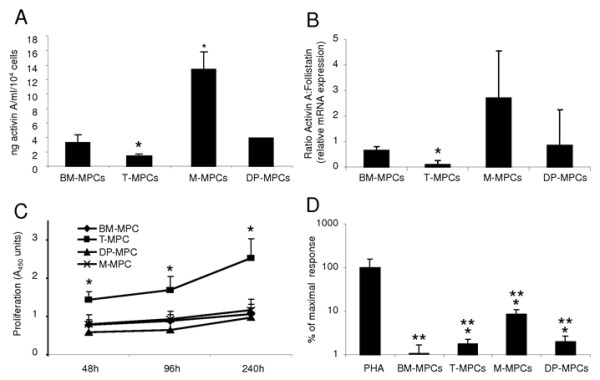
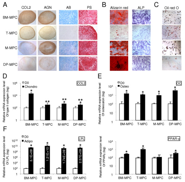
Methods: For this purpose, we directly compared MPCs isolated from different adult tissues, including bone marrow, tonsil, muscle, and dental pulp. We first examined and compared proliferation rates, immunomodulatory properties, and multidifferentiation potential of these MPCs in vitro. Next, we specifically evaluated activin A expression profile and activin A:follistatin ratio in MPCs from the four sources.
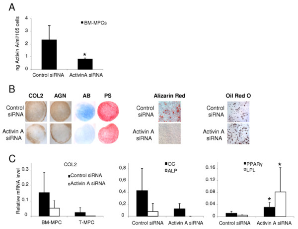
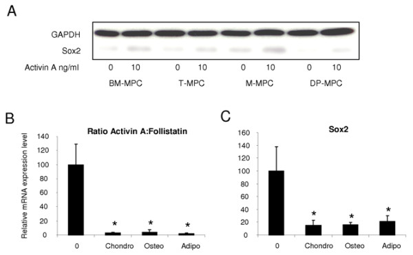
Results: The multidifferentiation potential of the MPCs is correlated with activin A level and/or the activin A:follistatin ratio. Interestingly, by siRNA-mediated activin A knockdown, activin A was shown to be required for the chondrogenic and osteogenic differentiation of MPCs. These findings strongly suggest that activin A has a pivotal differentiation-related role in the early stages of chondrogenesis and osteogenesis while inhibiting adipogenesis of MPCs.
Conclusions: This comparative analysis of MPCs from different tissue sources also identifies bone marrow-derived MPCs as the most potent MPCs in terms of multilineage differentiation and immunosuppression, two key requirements in cell-based regenerative medicine. In addition, this study implicates the significance of activin A as a functional marker of MPC identity.
Figures
Similar articles
-
Human palatine tonsil: a new potential tissue source of multipotent mesenchymal progenitor cells.Arthritis Res Ther. 2008;10(4):R83. doi: 10.1186/ar2459. Epub 2008 Jul 28. Arthritis Res Ther. 2008. PMID: 18662393 Free PMC article.
-
Pentosan polysulfate binds to STRO-1+ mesenchymal progenitor cells, is internalized, and modifies gene expression: a novel approach of pre-programing stem cells for therapeutic application requiring their chondrogenesis.Stem Cell Res Ther. 2017 Dec 13;8(1):278. doi: 10.1186/s13287-017-0723-y. Stem Cell Res Ther. 2017. PMID: 29237492 Free PMC article.
-
Characterization and Immunomodulatory Effects of Canine Adipose Tissue- and Bone Marrow-Derived Mesenchymal Stromal Cells.PLoS One. 2016 Dec 1;11(12):e0167442. doi: 10.1371/journal.pone.0167442. eCollection 2016. PLoS One. 2016. PMID: 27907211 Free PMC article.
-
Tissue source determines the differentiation potentials of mesenchymal stem cells: a comparative study of human mesenchymal stem cells from bone marrow and adipose tissue.Stem Cell Res Ther. 2017 Dec 6;8(1):275. doi: 10.1186/s13287-017-0716-x. Stem Cell Res Ther. 2017. PMID: 29208029 Free PMC article.
-
Characterisation of mesenchymal stromal cells in clinical trial reports: analysis of published descriptors.Stem Cell Res Ther. 2021 Jun 22;12(1):360. doi: 10.1186/s13287-021-02435-1. Stem Cell Res Ther. 2021. PMID: 34158116 Free PMC article. Review.
Cited by
-
Stem cells from human apical papilla decrease neuro-inflammation and stimulate oligodendrocyte progenitor differentiation via activin-A secretion.Cell Mol Life Sci. 2018 Aug;75(15):2843-2856. doi: 10.1007/s00018-018-2764-5. Epub 2018 Feb 7. Cell Mol Life Sci. 2018. PMID: 29417177 Free PMC article.
-
Osteochondral Tissue Chip Derived From iPSCs: Modeling OA Pathologies and Testing Drugs.Front Bioeng Biotechnol. 2019 Dec 17;7:411. doi: 10.3389/fbioe.2019.00411. eCollection 2019. Front Bioeng Biotechnol. 2019. PMID: 31921815 Free PMC article.
-
Tonsil-Derived Mesenchymal Stem Cells Differentiate into a Schwann Cell Phenotype and Promote Peripheral Nerve Regeneration.Int J Mol Sci. 2016 Nov 9;17(11):1867. doi: 10.3390/ijms17111867. Int J Mol Sci. 2016. PMID: 27834852 Free PMC article.
-
Differentiation and regeneration potential of mesenchymal progenitor cells derived from traumatized muscle tissue.J Cell Mol Med. 2011 Nov;15(11):2377-88. doi: 10.1111/j.1582-4934.2010.01225.x. J Cell Mol Med. 2011. PMID: 21129154 Free PMC article.
-
Direct differentiation of tonsillar biopsy-derived stem cells to the neuronal lineage.Cell Mol Biol Lett. 2021 Aug 18;26(1):38. doi: 10.1186/s11658-021-00279-4. Cell Mol Biol Lett. 2021. PMID: 34407767 Free PMC article.
References
-
- Noel D, Djouad F, Jorgense C. Regenerative medicine through mesenchymal stem cells for bone and cartilage repair. Curr Opin Investig Drugs. 2002;3:1000–1004. - PubMed
Publication types
MeSH terms
Substances
Grants and funding
LinkOut - more resources
Full Text Sources
Other Literature Sources
