Review
. 2010 Jul 16:11:68.
doi: 10.1186/1471-2156-11-68.

A symphony of inner ear developmental control genes

Affiliations
Review

A symphony of inner ear developmental control genes

Sumantra Chatterjee et al. BMC Genet. .

Abstract

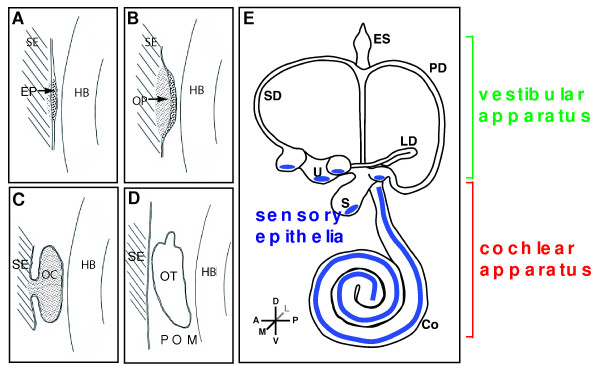
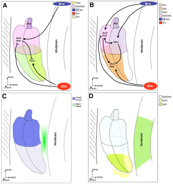
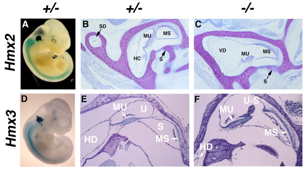
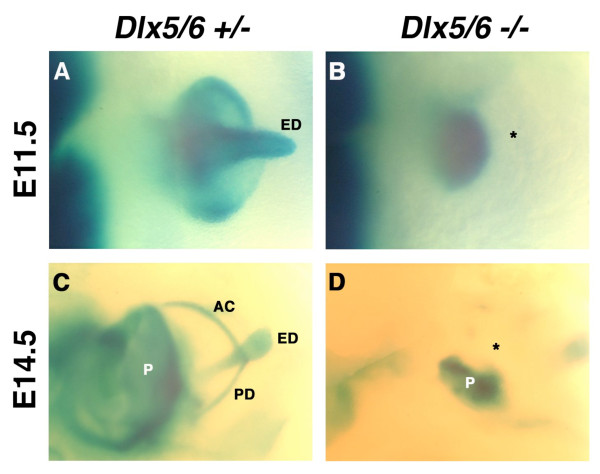
The inner ear is one of the most complex and detailed organs in the vertebrate body and provides us with the priceless ability to hear and perceive linear and angular acceleration (hence maintain balance). The development and morphogenesis of the inner ear from an ectodermal thickening into distinct auditory and vestibular components depends upon precise temporally and spatially coordinated gene expression patterns and well orchestrated signaling cascades within the otic vesicle and upon cellular movements and interactions with surrounding tissues. Gene loss of function analysis in mice has identified homeobox genes along with other transcription and secreted factors as crucial regulators of inner ear morphogenesis and development. While otic induction seems dependent upon fibroblast growth factors, morphogenesis of the otic vesicle into the distinct vestibular and auditory components appears to be clearly dependent upon the activities of a number of homeobox transcription factors. The Pax2 paired-homeobox gene is crucial for the specification of the ventral otic vesicle derived auditory structures and the Dlx5 and Dlx6 homeobox genes play a major role in specification of the dorsally derived vestibular structures. Some Micro RNAs have also been recently identified which play a crucial role in the inner ear formation.


Figures

Figure 1
Figure 1
Developmental milestones in mouse inner ear formation. Competence of surface ectoderm lateral to both sides of the hindbrain (HB) precedes any cell morphology changes. (A) Thickening of surface ectoderm (SE) to form the early placodes (EP) which is primarily driven by Fgf, Wnt and Pax genes. (B) Invagination of the otic placodes to form the otic pit (OP). (C) Further development and invagination of the otic pit to form the otic cup (OC) which pinches off from the surface ectoderm. (D) The separation from the overlying ectoderm gives rise to the otocyst (OT). (E) Subsequent morphogenesis to finalize the complex 3-dimensional labyrinth which is demarcated into vestibular and cochlear components. Sensory epithelia are shown in blue. Abbreviations: Co, cochlea; ES, endolymphatic sac; HB, hindbrain; LD, lateral semicircular duct; PD, posterior semicircular duct; POM, periotic mesenchyme; S, saccule; SD, superior semicircular duct; SE, surface ectoderm; U, utricle.
Figure 2
Figure 2
Representative expression patterns of genes controlling cochlear and vestibular specification. (A) Shh functions to maintain Pax2 and restrict Dlx5/Dlx6 in the medial wall of the otic vesicle in order to specify cochlear fate. Dlx5/Dlx6 specify the medial to dorsal most cells of the otic epithelium that give rise to the endolymphatic duct and vestibular apparatus. (B) Secretion of Shh from the notochord specifies the ventral most cells of the otic epithelium that express Otx1/Otx2 and possibly Pax2 which contribute to cochlear morphogenesis and outgrowth. In addition, Dlx5/Dlx6-dependent vestibular specifications and morphogenesis is dependent upon the activation of Gbx2 and Bmp4 function (not shown) and partial activation/expression of Otx1. Dlx5/Dlx6 also functions to restrict Pax2 expression to the medial wall of the otic vesicle epithelium. Thus, Dlx5/Dlx6 and Shh may functionally antagonize each other, through repression, to generate compartments of activities that specify the vestibular and cochlear cell fates. (C) Both Hmx2 and Hmx3 are required for cell fate determination and subsequent morphogenesis of the developing inner ear. Loss of both Hmx2 and Hmx3 results in the absence of the entire vestibular system. Msx1/Msx2 are expressed in the adjacent periotic mesenchyme and are critical for middle ear development. (D) Fgfs function with Shh in the periotic mesenchyme to initiate ventral otic capsule chondrogenesis via Brn4 and Tbx1 function (not shown). Fgfs are also expressed in the hindbrain epithelium adjacent to the otocyst and are important for induction of the otic placode.
Figure 3
Figure 3
Abnormal vestibular structure and morphogenesis in whole-mount β-galactosidase stained mid-gestation embryos lacking either Hmx2 or Hmx3. Early in development Hmx2 (A) is expressed throughout both the vestibular portions of the inner ear of heterozygous (A) embryos. Embryos that are homozygous for the absence of Hmx2 (C) have relatively normal cochlear development in the presence of severely dysmorphic vestibular development. The endolymphatic duct morphogenesis is retarded and the superior (SD), posterior (PD), and lateral or horizontal (HD) semicircular ducts appear to form a fused and primitive vestibular diverticulum (VD) and is associated with decreased maculae of the utricle (MU) and saccule (MS). In contrast, Hmx3 expression in the inner ear of heterozygous (D) and homozygous (F) embryos demonstrates expression throughout only the vestibular apparatus, including the ED and all three semicircular ducts. Embryos that are homozygous for the absence of Hmx3 (F) have mild faulty development of vestibular structures including a fusion of the utricular and saccular chambers (U-S) and a dysmorphic utricular maccula (MU) in the presence of circling behavior.
Figure 4
Figure 4
Abnormal vestibular morphogenesis in whole-mount β-galactosidase stained E11.5 and E14.5 embryos lacking both Dlx5 and Dlx6. Embryonic Dlx5 and Dlx6 expression in the inner ears of heterozygous (A, C) and homozygous (B, D) Dlx5/Dlx6 embryos demonstrates that vestibular morphogenesis is arrested by E11.5 with the absence of presumptive semicircular ducts and endolymphatic duct (ED, asterisk). At E14.5, Dlx5/Dlx6 expression normally defines the majority of the vestibular apparatus, including the anterior (AD), posterior (PD), and lateral (LD) semicircular ducts, ampullae (A) and ED. In contrast, the cell lineage of the presumptive vestibular apparatus is absent (asterisk) from Dlx5/Dlx6 null embryos, which develop a rudimentary pinna (P).

Similar articles

Cited by

References

    1. Raphael Y, Altschuler RA. Structure and innervation of the cochlea. Brain Res Bull. 2003;60:397–422. doi: 10.1016/S0361-9230(03)00047-9. - DOI - PubMed
    1. Takeuchi S, Ando M, Kakigi A. Mechanism generating endocochlear potential: role played by intermediate cells in stria vascularis. Biophys J. 2000;79:2572–82. doi: 10.1016/S0006-3495(00)76497-6. - DOI - PMC - PubMed
    1. Nance WE. The genetics of deafness. Ment Retard Dev Disabil Res Rev. 2003;9:109–19. doi: 10.1002/mrdd.10067. - DOI - PubMed
    1. Sando I, Orita Y, Miura M, Balaban CD. Vestibular abnormalities in congenital disorders. Ann N Y Acad Sci. 2001;942:15–24. - PubMed
    1. Walther C, Guenet JL, Simon D, Deutsch U, Jostes B, Goulding MD, Plachov D, Balling R, Gruss P. Pax: a murine multigene family of paired box-containing genes. Genomics. 1991;11:424–34. doi: 10.1016/0888-7543(91)90151-4. - DOI - PubMed

Substances