Comparative Study
. 2010 Nov;139(5):1642-53, 1653.e1-6.
doi: 10.1053/j.gastro.2010.07.008. Epub 2010 Jul 14.

Altered macrophage function contributes to colitis in mice defective in the phosphoinositide-3 kinase subunit p110δ

Affiliations
Comparative Study

Altered macrophage function contributes to colitis in mice defective in the phosphoinositide-3 kinase subunit p110δ

Jennifer K Uno et al. Gastroenterology. 2010 Nov.

Abstract

Background & aims: Innate immune responses are crucial for host defense against pathogens but need to be tightly regulated to prevent chronic inflammation. Initial characterization of mice with a targeted inactivating mutation in the p110δ subunit of phosphoinositide 3-kinase (PI3K p110δ(D910A/D910A)) revealed defects in B- and T-cell signaling and chronic colitis. Here, we further characterize features of inflammatory bowel diseases in these mice and investigate underlying innate immune defects.

Methods: Colons and macrophages from PI3K p110δ(D910A/D910A) mice were evaluated for colonic inflammation and innate immune dysfunction. Colonic p110δ messenger RNA expression was examined in interleukin (IL)-10(-/-) and wild-type germ-free mice during transition to a conventional microbiota. To assess polygenic impact on development of colitis, p110δ(D910A/D910A) mice were backcrossed to IL-10(-/-) mice.

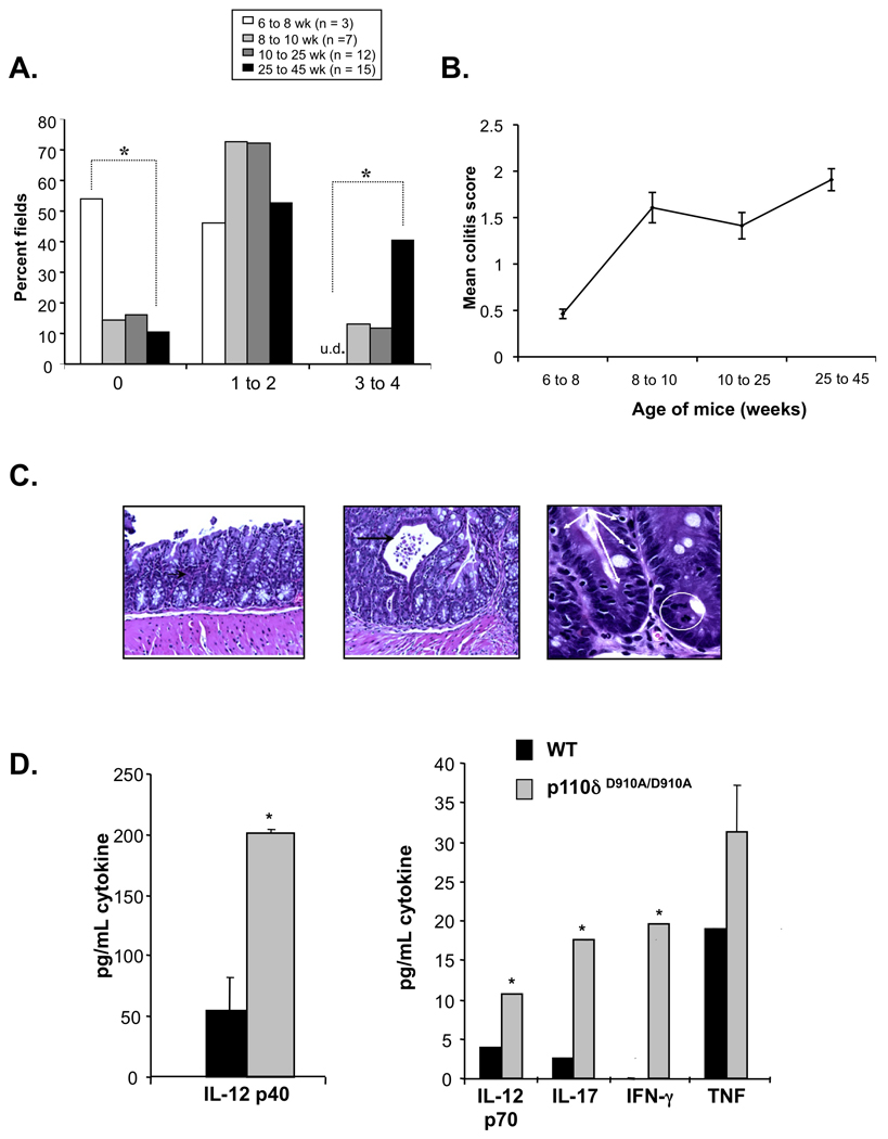
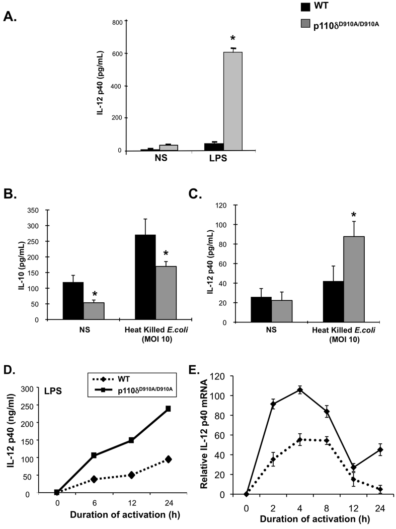
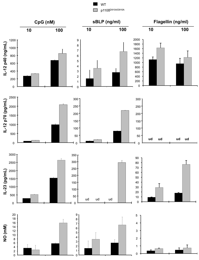
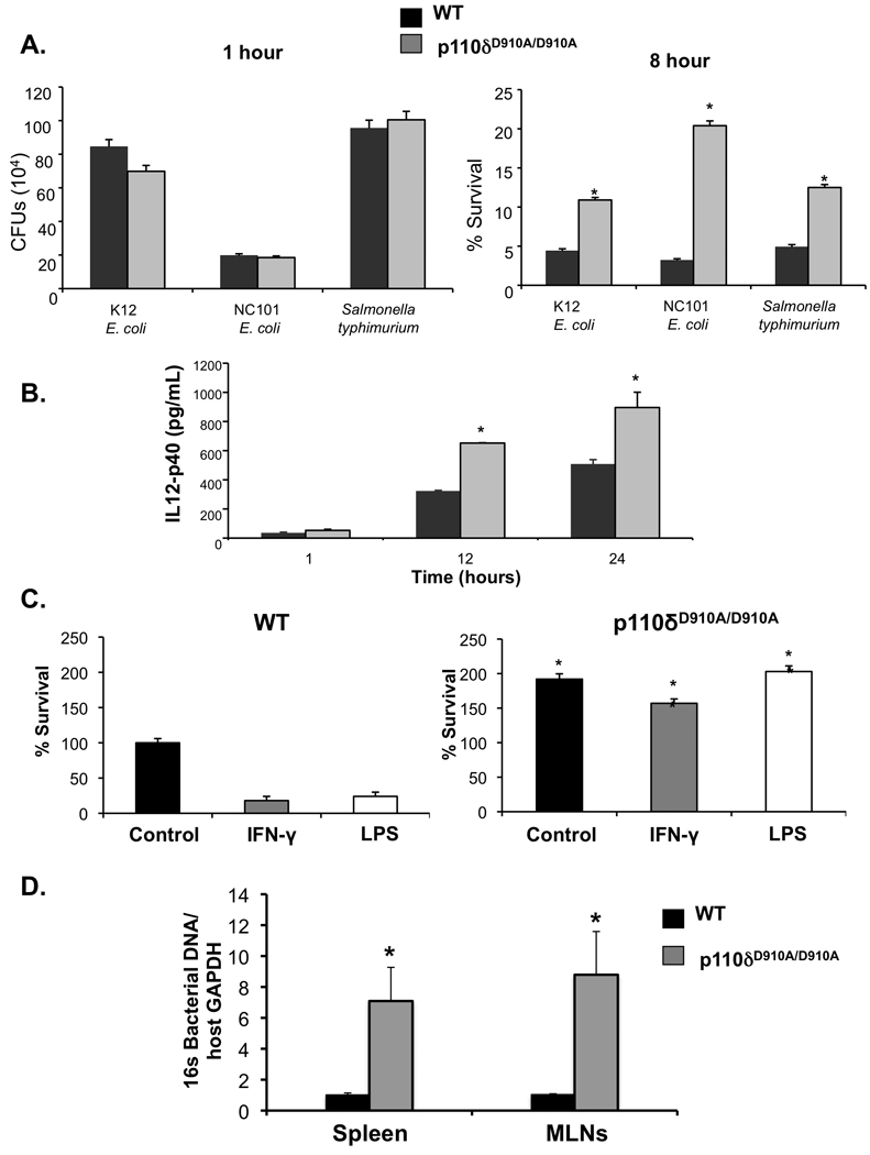
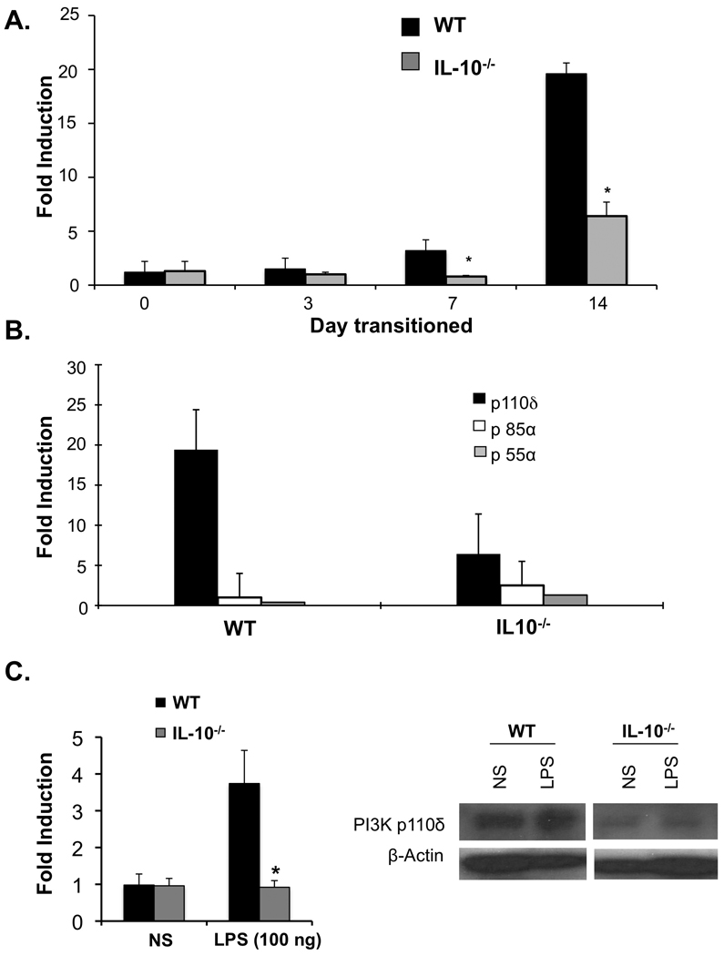
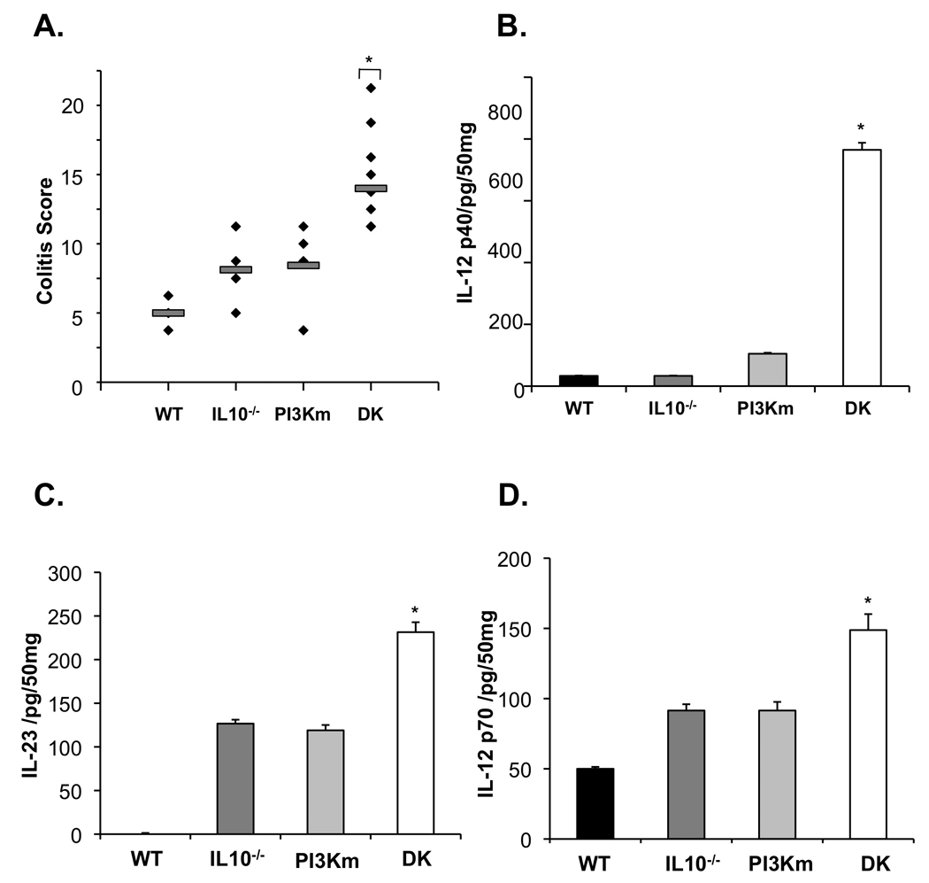
Results: A mild spontaneous colitis was shown in PI3K p110δ(D910A/D910A) mice at 8 weeks, with inflammation increasing with age. An inflammatory mucosal and systemic cytokine profile was characterized by expression of IL-12/23. In PI3K p110δ(D910A/D910A) macrophages, augmented toll-like receptor signaling and defective bactericidal activity were observed. Consistent with an important homeostatic role for PI3K p110δ, wild-type mice raised in a germ-free environment markedly up-regulated colonic PI3K p110δ expression with the introduction of the enteric microbiota; however, colitis-prone IL-10(-/-) mice did not. Moreover, PI3K p110δ(D910A/D910A) mice crossed to IL-10(-/-) mice developed severe colitis at an early age.

Conclusions: This study describes a novel model of experimental colitis that highlights the importance of PI3K p110δ in maintaining mucosal homeostasis and could provide insight into the pathogenesis of human inflammatory bowel disease.


Conflict of interest statement

Conflict of Interest: None of the authors listed above have any conflicts of interest to disclose.

Figures

Figure 1
Figure 1. PI3K p110δD910A/D910A mice develop colitis
(A and B) Histological scores of colonic sections from WT and PI3K p110δD910A/D910A mice at different ages. Results are represented as percentage of microscopic fields in each age group with score 0, 1 to 2 or 3 to 4 (A); or mean colitis scores (B). (*p<0.05 v. 25–45 WT week old mice) (C) Colonic sections from 10 week old PI3K p110dD910A/D910A mice demonstrate leukocytic infiltration of the lamina propria (white circle) and intraepithelial lymphocytes (white arrows) in the crypts. Focal crypt abscesses were observed (black arrow). (D) Colonic explants from WT (black bars) and PI3K p110δD910A/D910A (grey bars) mice were assayed for spontaneous secretion of cytokines. Error bars represent mean+SEM of 3 independent experiments.
Figure 2
Figure 2. PI3K p110δD910A/D910A mice display enhanced expression of IL-12 p40
(A) Splenocytes from WT (black bars) and PI3K p110δD910A/D910A (grey bars) mice were untreated (un) or stimulated with LPS (1 µg/ml) alone or LPS and IFN-γ (10 ng/ml) for 24 hours. IL-12 p40 was measured by ELISA. (B and C) Colonic macrophages from WT or PI3K p110dD910A/D910A mice were stimulated with heat killed E. coli (mode of multiplicity 10) for 24 hours. ELISAs were performed to assess IL-12 p40 (B) and IL-10 (C) levels. (D and E) Bone-marrow derived macrophages (BMMs) from WT (black bars) and PI3K p110δD910A/D910A (grey bars) mice were unstimulated (Un.) or stimulated with LPS (1 µg/ml) and supernatants analyzed for IL-12 p40 (D). PI3K p110dD910A/D910A BMMs were harvested at each time point and IL-12 p40 (Il12b) mRNA (E) levels were assessed by real-time RT-PCR. Results are expressed as fold induction normalized to β-actin. Error bars represent mean+SEM of 3 independent experiments (*p<0.05 v. WT cells).
Figure 3
Figure 3. PI3K p110δD910A/D910A macrophages demonstrate heightened sensitivity to TLR stimulation
BMMs from WT (black bars) and PI3K p110δD910A/D910A (grey bars) mice were stimulated with TLR9 (CpG), TLR2 (sBLP) or TLR5 (Flagellin) ligands for 24 hours. Supernatants were analyzed for IL-12 p40, IL-12 p70 or IL-23 secretion by ELISA and nitric oxide secretion by Greiss reaction. Error bars represent mean+SEM of 3 independent experiments.
Figure 4
Figure 4. PI3K p110δD910A/D910A macrophages demonstrate altered kinetics and magnitude of MAPK activation
(A) BMMs from WT or PI3K p110δD910A/D910A mice were stimulated with (right) LPS (100 ng/ml) or (left) sBLP (100 ng/ml) for the indicated periods of time and phosphorylation of Akt (p-Akt) was assayed by ELISA. Results are presented as a ratio of p-Akt to total Akt. (B) BMMs from WT and PI3K p110δD910A/D910A mice were stimulated with LPS (1 µg/ml) for the indicated times. Whole cell extracts were analyzed for phosphorylation of MAPK (JNK, ERK, p38) by Western blot. Results represent mean+SEM of 3 independent experiments (*p<0.05 vs WT).
Figure 5
Figure 5. PI3K p110δD910A/D910A BMMs demonstrate defective bactericidal activity
(A) WT and p110dD910A/D910A BMMs were cultured with K12 E. coli. NC101 E. coli or S. typhimurium. No significant bacterial recoveries were seen 1-hour post-bacterial infection (left panel). P13K p110δD910A/D910A BMMs demonstrate decreased bactericidal activity relative to wild type BMMs at 8 hours post-infection (right panel) (*p< 0.05 v WT BMMs). (B) IL-12 p40 production by ELISA was assessed in P13K p110dD910A/D910A BMMs infected with K12 E. coli. (C) BMMs were treated with LPS (100 ng/mL) or IFN-γ (10 ng/mL) prior to bacterial infection, and bacteria recovered from lysed cells 8 hours post-infection (*p< 0.05 vs WT BMMs). (D) Total bacterial DNA in spleen and mesenteric lymph nodes were detected by real-time PCR using primers for total 16S rRNA genes Primers for GAPDH were used to show loading control of host genomic DNA. Error bars represent mean+SEM of 3 independent experiments (*p < 0.05 vs. WT).
Figure 6
Figure 6. The enteric microbiota induces colonic PI3K p110δ expression in WT but not colitis-prone IL-10−/− mice
Germ-free (GF) WT and IL-10−/− mice were transitioned to a specific pathogen free (SPF) microbiota. Colonic mRNA was isolated and expression of PI3K p110δ, p85a, and p55a mRNA was assessed by real-time RT-PCR. (A) Colonic PI3K p110d was determined in WT and IL-10−/− mice at 0, 3, 7, and 14 days post colonization of GF mice with SPF microbiota (*p < 0.05 vs WT). (B) Colonic expression of PI3K p110δ, p85a, and p55a mRNA was examined 14 days post-transition of GF mice to SPF microbiota. Results are expressed as fold induction normalized to β-actin. Error bars represent mean+SEM of 3 independent experiments. (C) BMMs from WT and IL-10−/− mice were stimulated with LPS (100 ng) for the indicated times. PI3K p110δ mRNA levels were assessed by real-time RT-PCR (left panel). Results are expressed as fold induction normalized to β-actin and represent mean+SEM of 3 independent experiments (*p<0.05 vs WT). BMMs from WT and IL-10−/− mice were stimulated with LPS (100 ng) for 16 hours. Whole cell extracts were analyzed for PI3K p110δ by Western blot (right panel). Results are representative of three independent experiments.
Figure 7
Figure 7. IL -10−/− PI3K p110δD910A/D910A mice exhibit severe colitis at an early age
(A) Colitis scores were determined for 4-week-old IL-10−/−/PI3K p110δD910A/D910A (DK), WT, IL-10−/−, and PI3K p110δD910A/D910A (PI3Km) mice using criteria established for IL-10−/− mice by a pathologist blinded to genotype (*p < 0.05 vs WT). IL-12 p40 (C), IL-12 p70 (E) and IL-23 (D) protein in supernatants from colon explant cultures from IL-10−/−/PI3K p110δD910A/D910A (DK), WT, IL-10−/−, and PI3K p11δD910A/D910A (PI3Km) mice were analyzed by ELISA. Error bars represent mean+SEM of 3 independent experiments (*p< 0.05 vs. WT).

Comment in

References

    1. Xavier RJ, Podolsky DK. Unravelling the pathogenesis of inflammatory bowel disease. Nature. 2007;448:427–434. - PubMed
    1. Abreu MT. Toll-like receptor signalling in the intestinal epithelium: how bacterial recognition shapes intestinal function. Nat Rev Immunol. 10:131–144. - PubMed
    1. Rakoff-Nahoum S, Paglino J, Eslami-Varzaneh F, Edberg S, Medzhitov R. Recognition of commensal microflora by toll-like receptors is required for intestinal homeostasis. Cell. 2004;118:229–241. - PubMed
    1. Liew FY, Xu D, Brint EK, O'Neill LA. Negative regulation of toll-like receptor-mediated immune responses. Nat Rev Immunol. 2005;5:446–458. - PubMed
    1. Fukao T, Koyasu S. PI3K and negative regulation of TLR signaling. Trends Immunol. 2003;24:358–363. - PubMed

Publication types

MeSH terms