. 2010 Jul 23;142(2):203-17.
doi: 10.1016/j.cell.2010.06.001. Epub 2010 Jul 15.

SRD5A3 is required for converting polyprenol to dolichol and is mutated in a congenital glycosylation disorder

Affiliations

SRD5A3 is required for converting polyprenol to dolichol and is mutated in a congenital glycosylation disorder

Vincent Cantagrel et al. Cell. .

Abstract

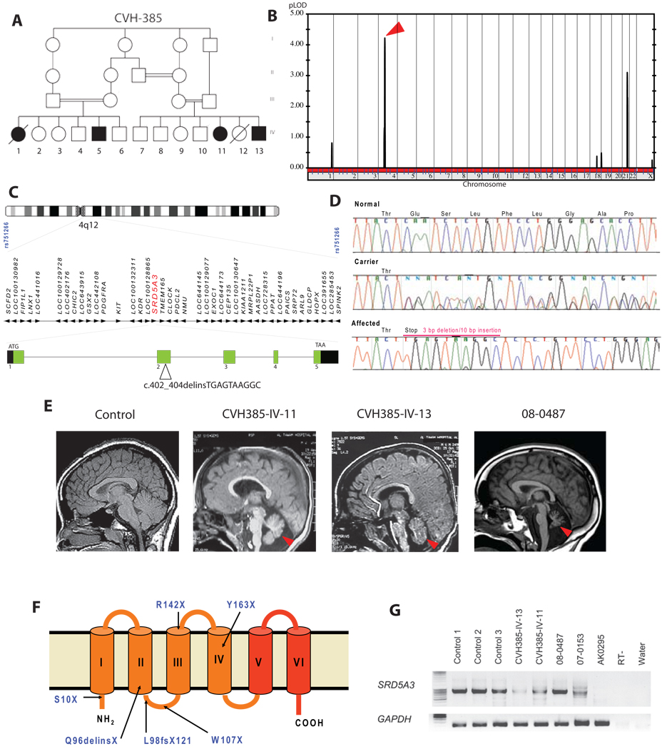
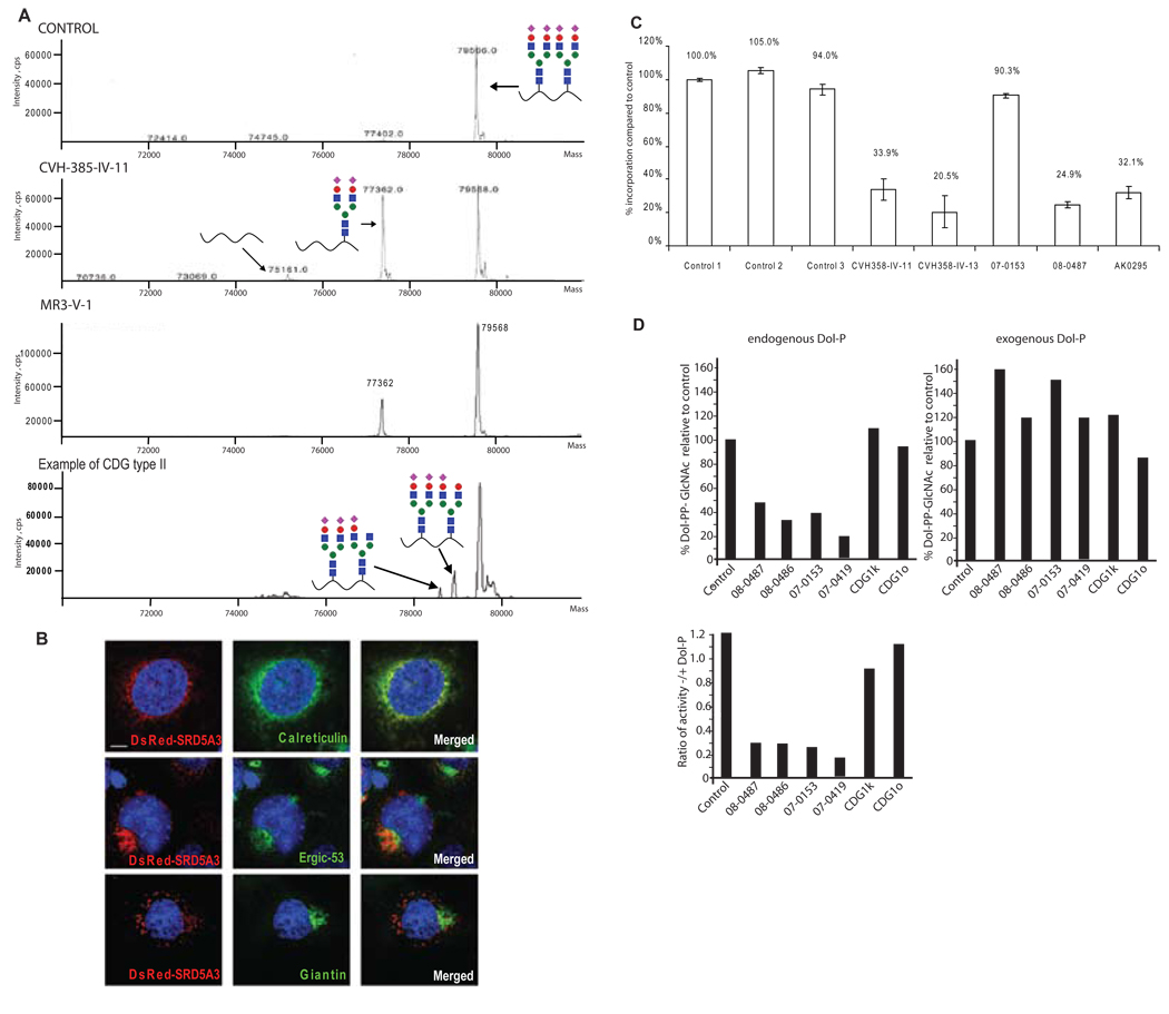
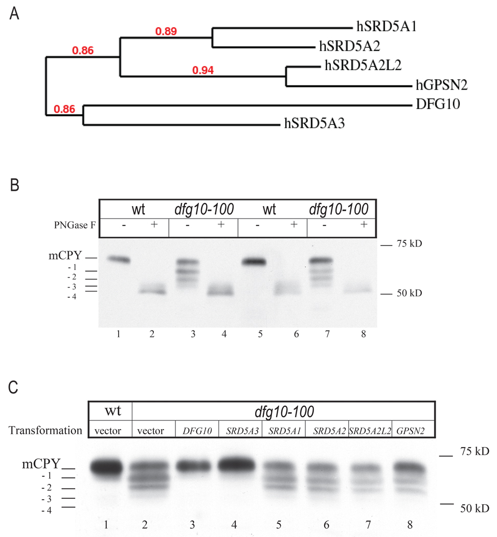
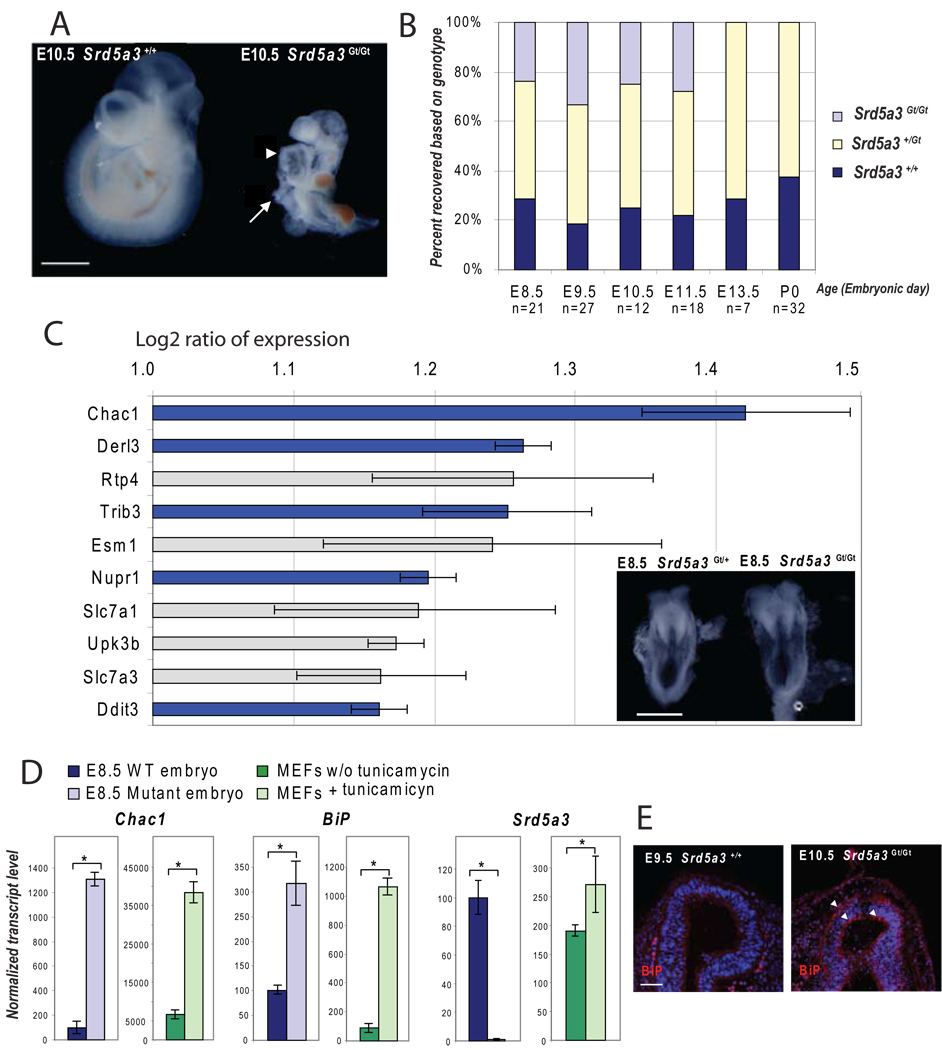
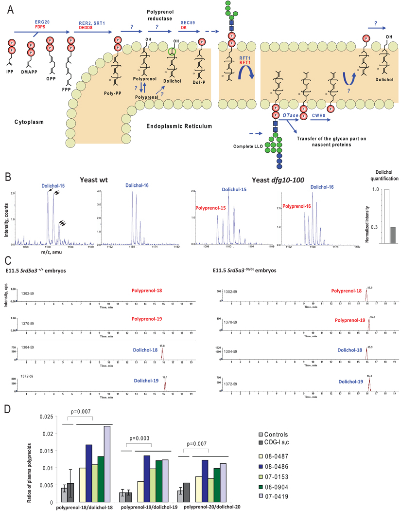
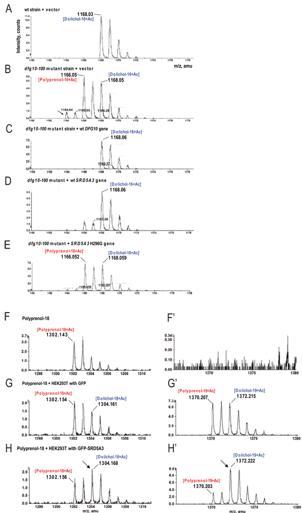
N-linked glycosylation is the most frequent modification of secreted and membrane-bound proteins in eukaryotic cells, disruption of which is the basis of the congenital disorders of glycosylation (CDGs). We describe a new type of CDG caused by mutations in the steroid 5alpha-reductase type 3 (SRD5A3) gene. Patients have mental retardation and ophthalmologic and cerebellar defects. We found that SRD5A3 is necessary for the reduction of the alpha-isoprene unit of polyprenols to form dolichols, required for synthesis of dolichol-linked monosaccharides, and the oligosaccharide precursor used for N-glycosylation. The presence of residual dolichol in cells depleted for this enzyme suggests the existence of an unexpected alternative pathway for dolichol de novo biosynthesis. Our results thus suggest that SRD5A3 is likely to be the long-sought polyprenol reductase and reveal the genetic basis of one of the earliest steps in protein N-linked glycosylation.


Figures

Figure 1
Figure 1. Identification of mutations in the SRD5A3 gene in patients with multisystemic syndrome including cerebellar hypoplasia
(A) Pedigree of family CVH-385 showing several levels of consanguinity with cousin marriages. The two branches each produced two affected offspring represented by filled symbols in generation IV. (B) Whole-genome analysis of linkage results with chromosomal position (x-axis) and multipoint LOD score (y-axis) showing a peak LOD score of 4.2 on chromosome 4 (arrowhead). (C) Expanded view of the candidate interval on chromosome 4q12, containing 42 candidate genes including SRD5A3 (red), spanning 25.5 kb of genomic DNA with 5 exons. A mutation in exon 2 was identified in family CVH-385. (D) DNA sequence of exon 2 of SRD5A3 from a control individual, an obligate carrier, and an affected family member from CVH-385. The mutation consists of a 3 bp deletion associated with a 10 bp insertion, resulting in a frame shift and premature termination at amino-acid 96 of 318, within the second of six transmembrane domains. (E) Brain MRI midline sagittal view showing cerebellar vermis hypoplasia (red arrowhead) in SRD5A3 mutated patients. (F) Topology model of SRD5A3 with mutations indicated and 6 transmembrane domains. Mutations were scattered throughout the ORF, all leading to predicted protein termination before the steroid reductase domain (in red). (G) Amplification of the SRD5A3 transcript by RT-PCR using RNA extracted from controls and patient fibroblasts. The expression level of the gene is lower in almost all patients CVH-385-IV-13, CVH-385-IV-11, 07–0153 compared to control, suggesting nonsense mediated mRNA decay. No expression was detected in patient AK0295, due to a homozygous genomic rearrangement. RT-, no reverse transcriptase; water, no cDNA. See also Figure S1.
Figure 2
Figure 2. SRD5A3 mutated patients have a congenital disorder of glycosylation type I caused by a defect in lipid-linked oligosaccharide (LLO) synthesis, rescued in vitro with exogenous dolichol phosphate
(A) Mass spectra of transferrin, normally N-glycosylated on two sites, Asn-432 and Asn-630 (control). Transferrin containing a single N-glycan in SRD5A3 patient samples was increased, indicating a CDG type I disorder. An example of transferrin profile from CDG type II patient, with two glycan chains but abnormal structure (depicted by lack of certain sugar moieties) for comparison. (B) Intra cellular localization of SRD5A3 containing a N-terminus DsRed tag (center panel) in COS7 cells costained with antibody against ER specific marker, calreticulin, ERGIC specific marker ERGIC53 and Golgi specific marker Giantin. DsRed-SRD5A3 colocalized with most of the ER whereas Giantin staining did not colocalize. Scale bar = 10µm. (C) Incorporation of [3H]-mannose into LLO after labeling of human fibroblasts. The results indicate severely reduced levels of LLO in 4 of 5 patient samples. (D) Rescue of LLO precursor levels with exogenous Dol-P. GlcNAc transferase activity in fibroblasts was measured. Microsomal fractions from fibroblasts were incubated with radioactive GlcNAc and then Dol-PP-GlcNAc1/2 formation was analyzed by TLC. Extracts from patients’ fibroblasts produced a reduced amount of Dol-PP-GlcNAc1/2. However the addition of exogenous Dol-P rescued this defect. See also Figure S2.
Figure 3
Figure 3. N-glycosylation phenotype of dfg10–100 yeast mutant and rescue with human SRD5A3
(A) Phylogenic tree representation of the yeast protein DFG10 and human proteins presenting a steroid 5-alpha reductase domain, with branch support value indicated in red. (B) N-glycosylation status of the yeast protein CPY in yeast wt and dfg10–100 mutant strain, mutated by transposon insertion. Two colonies from each strain were tested. CPY is post-translationally modified by the addition of four glycan chains. In the dfg10–100 mutants, a protein lacking one, two or tree glycan chains is detected. Protein extracts were treated with PNGaseF to remove the N-glycans. (C) Glycosylation status of CPY in dfg10–100 mutant transformed with each steroid 5-alpha reductase domain containing gene from human. Only SRD5A3 showed rescue effect. Positions of mature CPY (mCPY) and the different glycoforms (−1,−2,−3,−4) are indicated. Vector indicates empty vector control. See also Figure S3.
Figure 4
Figure 4. Characterization of homozygous Srd5a3Gt/Gt gene trap mouse embryos
(A) Phenotype at E10.5 shows failure to rotate, ventral body wall defect (arrow) and dilated heart (arrowhead). Scale bar 1 mm. (B) Graphic representation of the genotype obtained from the progeny of heterozygous mating, with lethality appearing between E11.5 and E13.5. (C) Genes overexpressed in Srd5a3Gt/Gt at E8.5 detected using 44k mouse genome oligo microarray (1 is the Log 2 of a 2 fold expression increase, error bars represent the mean +/− standard deviation from 4 independent experiments). Among 10 of the most upregulated genes, 5 (in blue) are involved in the unfolded protein response pathway UPR). Morphology of heterozygous and homozygous Srd5a3 mutant embryos at E8.5, before embryo axial rotation. Scale bar 500 µm. (D) Real-time RT-PCR confirming activation of the UPR pathway (Errors bars are means +/− standard deviations, asterisks indicate p<0.05, n=3). (E) Immunofluorescence staining showing expression of BiP protein (red), a marker of the UPR pathway activation, in the neuroepithelium of the forebrain vesicle (arrowheads). Scale bar 50µm. See also Figure S4.
Figure 5
Figure 5. Analysis of polyprenols and dolichols, in yeast, mouse and human, using LC-MS
(A) De novo biosynthesis and recycling of dolichol in eukaryotic cells with the yeast (in blue) and human (in red) enzymes involved. Isopentenylpyrophosphate (IPP) is the building block for all polyprenoids. IPP molecules are added sequentially in trans configuration, on dimethylallyl pyrophosphate (DMAPP) via the farnesyl pyrophosphate synthase (ERG20/FDPS) to form geranyl pyrophosphate (GPP) and then farnesyl pyrophosphate (FPP). More IPP units are then added in cis-configuration on FPP by the cis-prenyl transferases (RER2, SRT1/DHDDS), producing long polyprenoids that are embedded in the ER membrane. Once the final length is reached, both phosphate residues are released by unidentified phosphatases. The alpha-isoprene unit of the polyprenol is subsequently reduced by an NADPH-dependent microsomal reductase. For this step, the corresponding aldehydes have also been suggested as intermediates (Sagami et al., 1996). Finally the dolichol-specific kinase (SEC59/DK) transfers a phosphate from CTP to dolichol. Dol-P is used to build the lipid linked oligosaccharide (LLO). Once the oligosaccharide structure is transferred to specific asparagine residues, Dol-P is release on the luminal leaflet of the ER and dephosphorylated by a pyrophosphatase (CWH8). (B) Mass spectra of Dolichol-15,16 in wt and dfg10–100 yeast strains showing accumulation of corresponding polyprenols in the mutant strain. Single, double and triple arrows highlight natural isotopic distribution, each offset by one atomic mass unit (amu). Polyprenol was detected only in mutant (red), by identification of a spectrum that partially overlapped with dolichol. Quantification of the dolichol content of yeast mutant indicates 70% reduction (following subtraction of polyprenol isotopic contribution, see methods, white bar = wt, grey bar = mutant). (C) Scans of Dolichol-18,19 in wt and E11.5 Srd5a3Gt/Gt embryos showing the accumulation of corresponding polyprenols in the mutant embryos. (D) Ratio of polyprenol-18,19,20 on dolichol-18,19,20 calculated with lipid plasma level from control and SRD5A3 mutated patients, showing a significant increase of these ratios compared to controls (n=10) and other types of CDG (n=4). Error bars represent the mean +/− standard deviation. Two-tailed student’s t-test was used for statistical analysis. Color bars represent individual measurement for 5 patients with SRD5A3 mutation. Error bar was not generated for pol-20/dol-20 ratio in the group CDGI-a,c as polyprenol levels were undetectable in 3 of 4 patients.
Figure 6
Figure 6. In vivo and in vitro polyprenol reduction promoting activity of SRD5A3
(A–E) LC-MS analysis of lipid extract from yeast cultured in minimal media. (A) Only dolichol is detected in wt yeast strains transformed with pYX212 empty vector. (B) In dfg10–100 strain transformed with pYX212 empty vector, accumulation of polyprenol relative dolichol is evident. An additional compound (arrows) was tentatively identified as polyprenal, previously suggested as an intermediate in yeast during in vitro dolichol biosynthesis (Sagami et al., 1996). (C) In the dfg10–100 strain transformed with wt DFG10 gene, no polyprenol accumulation was detected. (D) Transformation of the dfg10–100 strain with the human SRD5A3 gene corrects polyprenol accumulation, although low levels are still detected. (E) Transformation of the dfg10–100 strain with the human SRD5A3 enzymatically null H296G mutation fails to correct the polyprenol accumulation. (F–H) LC-MS analysis of a lipid extract from an in vitro experiment performed with the exogenous substrate, polyprenol-18, in which cell lysates were used as the source of enzyme. (F,F’) Polyprenol-18 spectrum after incubation in the reaction buffer without cell lysate. No polyprenol-19 or any forms of dolichol are evident. (G,G’) After incubation with HEK293T cell lysate transfected with GFP, part of polyprenol-18 is elongated in polyprenol-19 and 28% is reduced to the corresponding dolichol. (H,H’) In presence of lysate from cell over-expressing GFP-SRD5A3, 67% of the initial polyprenol is reduced to dolichol (arrows). Both cell lysates show similarly elongation of polyprenols. See also Figure S5 and Figure S6.

Comment in

Similar articles

Cited by

References

    1. Aebi M, Hennet T. Congenital disorders of glycosylation: genetic model systems lead the way. Trends Cell Biol. 2001;11:136–141. - PubMed
    1. Aggarwal S, Thareja S, Verma A, Bhardwaj TR, Kumar M. An overview on 5alpha-reductase inhibitors. Steroids. 2009 - PubMed
    1. Al-Gazali L, Hertecant J, Algawi K, El Teraifi H, Dattani M. A new autosomal recessive syndrome of ocular colobomas, ichthyosis, brain malformations and endocrine abnormalities in an inbred Emirati family. Am J Med Genet A. 2008;146:813–819. - PubMed
    1. Alber T, Kawasaki G. Nucleotide sequence of the triose phosphate isomerase gene of Saccharomyces cerevisiae. J Mol Appl Genet. 1982;1:419–434. - PubMed
    1. Andersson S, Berman DM, Jenkins EP, Russell DW. Deletion of steroid 5 alpha-reductase 2 gene in male pseudohermaphroditism. Nature. 1991;354:159–161. - PMC - PubMed

Publication types

MeSH terms