. 2010 Sep 15;247(3):169-78.
doi: 10.1016/j.taap.2010.07.004. Epub 2010 Jul 15.

Role of caspase-1 and interleukin-1beta in acetaminophen-induced hepatic inflammation and liver injury

Affiliations

Role of caspase-1 and interleukin-1beta in acetaminophen-induced hepatic inflammation and liver injury

C David Williams et al. Toxicol Appl Pharmacol. .

Abstract

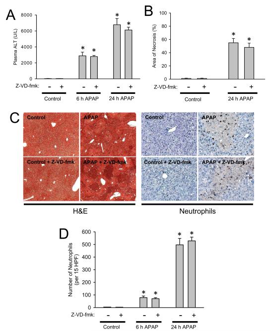
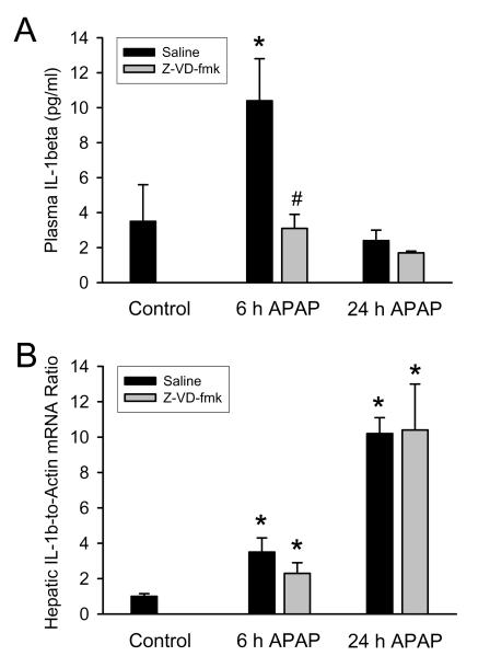
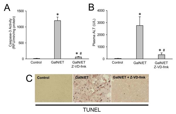
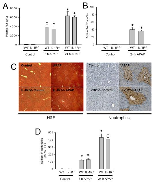
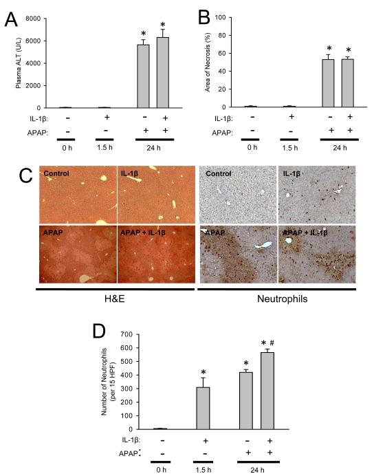
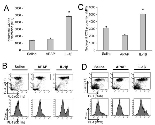
Acetaminophen (APAP) overdose can result in serious liver injury and potentially death. Toxicity is dependent on metabolism of APAP to a reactive metabolite initiating a cascade of intracellular events resulting in hepatocellular necrosis. This early injury triggers a sterile inflammatory response with formation of cytokines and innate immune cell infiltration in the liver. Recently, IL-1beta signaling has been implicated in the potentiation of APAP-induced liver injury. To test if IL-1beta formation through caspase-1 is critical for the pathophysiology, C57Bl/6 mice were treated with the pan-caspase inhibitor Z-VD-fmk to block the inflammasome-mediated maturation of IL-1beta during APAP overdose (300 mg/kg APAP). This intervention did not affect IL-1beta gene transcription but prevented the increase in IL-1beta plasma levels. However, APAP-induced liver injury and neutrophil infiltration were not affected. Similarly, liver injury and the hepatic neutrophilic inflammation were not attenuated in IL-1-receptor-1 deficient mice compared to wild-type animals. To evaluate the potential of IL-1beta to increase injury, mice were given pharmacological doses of IL-1beta after APAP overdose. Despite increased systemic activation of neutrophils and recruitment into the liver, there was no alteration in injury. We conclude that endogenous IL-1beta formation after APAP overdose is insufficient to activate and recruit neutrophils into the liver or cause liver injury. Even high pharmacological doses of IL-1beta, which induce hepatic neutrophil accumulation and activation, do not enhance APAP-induced liver injury. Thus, IL-1 signaling is irrelevant for APAP hepatotoxicity. The inflammatory cascade is a less important therapeutic target than intracellular signaling pathways to attenuate APAP-induced liver injury.


Figures

Figure 1
Figure 1
Acetaminophen-induced liver injury in C57Bl/6 mice with or without caspase inhibitor. Animals were first treated with 300 mg/kg APAP and then two hours later with 10 mg/kg Z-VD-fmk or vehicle control. (A) Plasma ALT at 6 h and 24 h; (B) area of necrosis at 24 h. (C) Representative H&E-stained liver sections (x50 magnification) and immunohistochemistry for hepatic neutrophil accumulation (x200 magnification) are shown for controls and animals treated with APAP for 24 h. (D) Neutrophil numbers were quantified in 15 randomly selected high power fields (HPF; x400). Data represent means ± SE of n = 5 animals per group. *P<0.05 (compared to untreated controls).
Figure 2
Figure 2
Interleukin-1β (IL-1β) protein levels in plasma (A) and hepatic IL-1β mRNA levels (B) were measured 6 and 24 h after administration of 300 mg/kg acetaminophen (APAP) in C57Bl/6 mice with or without the pan-caspase inhibitor (Z-VD-fmk). mRNA levels are expressed as IL-1β mRNA-to-β-actin mRNA ratio. The values of untreated controls were set as 1 and the fold change of the treated animals was calculated. Data represent means ± SE of n = 4-5 animals per group. *P<0.05 (compared to untreated control). #P<0.05 (compared to APAP-only equivalent time point)
Figure 3
Figure 3
Effects of 10 mg/kg Z-VD-fmk on galactosamine/endotoxin (GalN/ET)-induced liver injury. Animals were treated with saline or 700 mg/kg GalN and 100 μg/kg ET and then three hours later with 10 mg/kg Z-VD-fmk or vehicle control. (A) Caspase-3 activity at 6 h; (B) Plasma ALT at 6 h. (C) Representative TUNEL staining of animals from each group. Data represent means ± SE of n = 3 animals per group. *P<0.05 (compared to saline-treated controls)
Figure 4
Figure 4
Acetaminophen-induced liver injury in C57Bl/6 wild-type mice or IL-1R1-deficient mice. Animals were treated with 300 mg/kg APAP. (A) Plasma ALT at 6 h and 24 h; (B) area of necrosis at 24 h. (C) Representative H&E-stained liver sections (x50 magnification) and immunohistochemistry for hepatic neutrophil accumulation (x200 magnification) are shown for control and APAP-treated mice at 24 h; (D) Neutrophil numbers were quantified in 15 randomly selected high power fields (HPF; x400). Data represent means ± SE of n = 6 animals per group. *P<0.05 (compared to untreated controls).
Figure 5
Figure 5
Acetaminophen-induced liver injury in C57Bl/6 mice with or without IL-1β treatment. Mice were treated with 300 mg/kg APAP and/or 20 μg/kg IL-1β or vehicle. (A) Plasma ALT levels at 24 h; (B) area of necrosis at 24 h; (C) Representative H&E-stained liver sections (x50 magnification) and immunohistochemistry for hepatic neutrophil accumulation (x200 magnification) are shown for control and treated mice. (D) Neutrophil numbers were quantified in 15 randomly selected high power fields (HPF; x400). Data represent means ± SE of n = 6 animals per group. *P<0.05 (compared to untreated controls). #P<0.05 (compared to APAP only).
Figure 6
Figure 6
Neutrophil priming in peripheral blood. C57Bl/6 mice (n=4 per group) were treated with 20 mL/kg saline (6 h), 300 mg/kg APAP (6 h), or 20 μg/kg IL-1β (1.5 h). Immediately after in vivo stimulation whole blood was stained for CD11b and Gr-1 surface expression and neutrophils (Gr-1bright cells) were analyzed by flow cytometry. (A) The mean CD11b fluorescent intensities of each treatment group are shown and (B) representative CD11b surface-expression dot plots and histograms for saline, APAP and IL-1β. To determine reactive oxygen species (ROS) priming after in vivo treatment, whole blood was stimulated ex vivo with PMA or saline. Upon PMA-induced ROS production DHR-123 is converted to R-123 and quantified in neutrophils by flow cytometry. (C) The mean ROS fluorescent intensities of each treatment group are shown and (D) representative ROS dot plots and histograms for saline, APAP and IL-1β. Data represent means ± SE of n = 4 animals per group. *P<0.05 (compared to saline-treated controls).

References

    1. Bajt ML, Farhood A, Jaeschke H. Effects of CXC chemokines on neutrophil activation and sequestration in hepatic vasculature. Am. J. Physiol. Gastrointest. Liver Physiol. 2001;281:G1188–G1195. - PubMed
    1. Bajt ML, Yan HM, Farhood A, Jaeschke H. Plasminogen activator inhibitor-1 limits liver injury and facilitates regeneration after acetaminophen overdose. Toxicol. Sci. 2008;104:419–427. - PMC - PubMed
    1. Blazka ME, Elwell MR, Holladay SD, Wilson RE, Luster MI. Histopathology of acetaminophen-induced liver changes: role of interleukin 1 alpha and tumor necrosis factor alpha. Toxicol. Pathol. 1996;24:181–189. - PubMed
    1. Blazka ME, Wilmer JL, Holladay SD, Wilson RE, Luster MI. Role of proinflammatory cytokines in acetaminophen hepatotoxicity. Toxicol. Appl. Pharmacol. 1995;133:43–52. - PubMed
    1. Boess F, Bopst M, Althaus R, Polsky S, Cohen SD, Eugster HP, Boelsterli UA. Acetaminophen hepatotoxicity in tumor necrosis factor/lymphotoxin-alpha gene knockout mice. Hepatology. 1998;27:1021–1029. - PubMed

Publication types

MeSH terms