. 2010 Dec;172(3):270-5.
doi: 10.1016/j.jsb.2010.07.003. Epub 2010 Jul 15.

Keratin homogeneity in the tail feathers of Pavo cristatus and Pavo cristatus mut. alba

Affiliations

Keratin homogeneity in the tail feathers of Pavo cristatus and Pavo cristatus mut. alba

S Pabisch et al. J Struct Biol. 2010 Dec.

Abstract

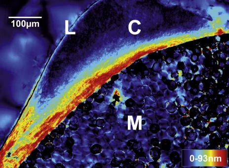
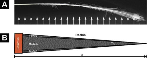
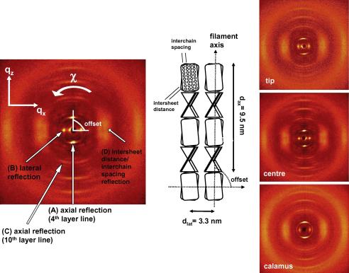
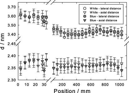
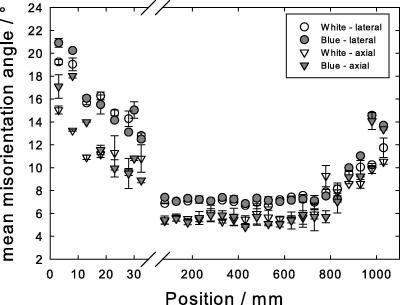
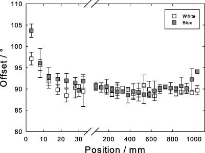
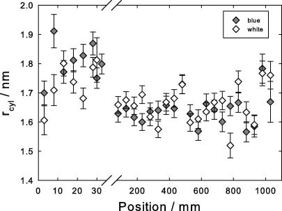
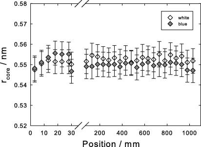
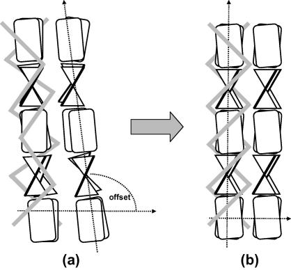
The keratin structure in the cortex of peacocks' feathers is studied by X-ray diffraction along the feather, from the calamus to the tip. It changes considerably over the first 5 cm close to the calamus and remains constant for about 1m along the length of the feather. Close to the tip, the structure loses its high degree of order. We attribute the X-ray patterns to a shrinkage of a cylindrical arrangement of β-sheets, which is not fully formed initially. In the final structure, the crystalline beta-cores are fixed by the rest of the keratin molecule. The hydrophobic residues of the beta-core are locked into a zip-like arrangement. Structurally there is no difference between the blue and the white bird.


Figures

Fig. 1
Fig. 1
Polscope (Cri Abrio System, L.O.T. Oriel, Germany) image of a rachis cross-section. Below the diagonal the foamy medulla structure (M) can be seen. It is surrounded by the cortex (C) of varying thickness, appearing moon-shaped. This is sub-structured into layers. Towards the top left, the cortex is covered by protective, presumably lipid, layer (L). Birefringent retardation (0–93 nm) is color coded.
Fig. 2
Fig. 2
(A) The tail cover feather of Pavo cristatus sub. alba subjected to X-ray investigation. The arrows indicate approximately the multiple beam positions. (B) Sketch of the conical shape of the rachis. Both outer diameter and cortex thickness decrease linearly from the calamus to the tip, forming a self-similar cone filled by the medulla. Diffraction patterns were taken from the cortex only.
Fig. 3
Fig. 3
The four reflections analyzed, (A), (B), (C), (D), are indicated on a typical diffraction pattern, left image. Corresponding real space arrangement proposed by Fraser and Parry (1996) for avian keratin is visible in the center. Right images, three typical scattering patterns along the length of a white peacock feather, from the regions close to the calamus, the center and close to the tip.
Fig. 4
Fig. 4
Positional change of the axial fourth layer line reflection (A – triangles) and the first lateral reflection (B – circles) along the length of the rachis of the feather: open symbols, white peacock, gray symbols, blue peacock. Note that the abscissa is broken to allow for the strong change in the region close to the calamus, positions 0–30 mm.
Fig. 5
Fig. 5
Mean misorientation angle along the length of the feather, i.e. the Gaussian half-width of the azimuthal spread of the axial reflection from the axial fourth layer line, (A – triangles), and the first lateral reflection, (B – circles). Open symbols, white peacock, gray symbols, blue peacock. The abscissa is broken to allow for the strong change in the region close to the calamus, positions 0–30 mm.
Fig. 6
Fig. 6
Offset angle between the axial reflection (A) (length direction of keratin filament) and the lateral reflection (B) (perpendicular direction of the keratin filament). Open symbols, white peacock, gray symbols, blue peacock. The abscissa is broken to allow for the strong change in the region close to the calamus, positions 0–30 mm.
Fig. 7
Fig. 7
Cylinder radius evaluated from the axial 10th layer line (C). Open symbols, white peacock, gray symbols, blue peacock. Note the expanded scale near the calamus, positions 0–30 mm.
Fig. 8
Fig. 8
Core radius rcore (half the value of the intersheet distance) from the broad lateral reflection (D). Open symbols, white peacock, gray symbols, blue peacock. Note the expanded scale near the calamus, positions 0–30 mm.
Fig. 9
Fig. 9
Structural change from the calamus to the central region: close to the calamus, (a), the β-sheets are misoriented with respect to the filament axis. Away from the calamus, (b), they are increasingly aligned and form a highly oriented structure with filament axis and lateral packing being perfectly perpendicular. The grey bars symbolize the outer parts of the molecule, which are not organized in pleated sheets. Both the diameter of the filaments and their distance decreases.

References

    1. Alexander D.E., Gong E., Martin L.D., Burnham D.A., Falk A.R. Model tests of gliding with different hindwing configurations in the four-winged dromaeosaurid Microraptor gui. PNAS. 2010;107:2972–2976. - PMC - PubMed
    1. Astbury W.T., Beighton E. Structure of feather keratin. Nature. 1961;191:171–173.
    1. Astbury W.T., Marwick T.C. X-ray interpretation of the molecular structure of feather keratin. Nature. 1932;130:309–310.
    1. Astbury W.T., Woods H.J. X-ray studies of the structure of hair, wool, and related fibres. II. The molecular structure and elastic properties of hair keratin. Philosophical Transactions of the Royal Society of London, Series A. 1934;232:333–394.
    1. Benton M.J. Evolutionary biology: new take on the red queen. Nature. 2010;463:306–307. - PubMed

Publication types

LinkOut - more resources