A superior semicircular canal dehiscence-induced air-bone gap in chinchilla
- PMID: 20638462
- PMCID: PMC2936693
- DOI: 10.1016/j.heares.2010.07.002
A superior semicircular canal dehiscence-induced air-bone gap in chinchilla
Abstract
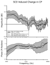
An SCD is a pathologic hole (or dehiscence) in the bone separating the superior semicircular canal from the cranial cavity that has been associated with a conductive hearing loss in patients with SCD syndrome. The conductive loss is defined by an audiometrically determined air-bone gap that results from the combination of a decrease in sensitivity to air-conducted sound and an increase in sensitivity to bone-conducted sound. Our goal is to demonstrate, through physiological measurements in an animal model, that mechanically altering the superior semicircular canal (SC) by introducing a hole (dehiscence) is sufficient to cause such an air-bone gap. We surgically introduced holes into the SC of chinchilla ears and evaluated auditory sensitivity (cochlear potential) in response to both air- and bone-conducted stimuli. The introduction of the SC hole led to a low-frequency (<2000 Hz) decrease in sensitivity to air-conducted stimuli and a low-frequency (<1000 Hz) increase in sensitivity to bone-conducted stimuli resulting in an air-bone gap. This result was consistent and reversible. The air-bone gaps in the animal results are qualitatively consistent with findings in patients with SCD syndrome.
Figures


Similar articles
-
The effect of superior canal dehiscence on cochlear potential in response to air-conducted stimuli in chinchilla.Hear Res. 2005 Dec;210(1-2):53-62. doi: 10.1016/j.heares.2005.07.003. Epub 2005 Sep 8. Hear Res. 2005. PMID: 16150562 Free PMC article.
-
Clinical, experimental, and theoretical investigations of the effect of superior semicircular canal dehiscence on hearing mechanisms.Otol Neurotol. 2004 May;25(3):323-32. doi: 10.1097/00129492-200405000-00021. Otol Neurotol. 2004. PMID: 15129113
-
A third window of the posterior semicircular canal: an animal model.Laryngoscope. 2010 May;120(5):1034-7. doi: 10.1002/lary.20831. Laryngoscope. 2010. PMID: 20422700
-
Quantification of hearing loss in patients with posterior semicircular canal dehiscence.Acta Otolaryngol. 2015;135(10):974-7. doi: 10.3109/00016489.2015.1060630. Epub 2015 Jun 24. Acta Otolaryngol. 2015. PMID: 26107020 Review.
-
Canal dehiscence.Curr Opin Neurol. 2011 Feb;24(1):25-31. doi: 10.1097/WCO.0b013e328341ef88. Curr Opin Neurol. 2011. PMID: 21124219 Review.
Cited by
-
Semicircular canal biomechanics in health and disease.J Neurophysiol. 2019 Mar 1;121(3):732-755. doi: 10.1152/jn.00708.2018. Epub 2018 Dec 19. J Neurophysiol. 2019. PMID: 30565972 Free PMC article. Review.
-
Superior Canal Dehiscence Similarly Affects Cochlear Pressures in Temporal Bones and Audiograms in Patients.Ear Hear. 2020 Jul/Aug;41(4):804-810. doi: 10.1097/AUD.0000000000000799. Ear Hear. 2020. PMID: 31688316 Free PMC article.
-
Mechanisms of Hearing Loss in a Guinea Pig Model of Superior Semicircular Canal Dehiscence.Neural Plast. 2018 Apr 24;2018:1258341. doi: 10.1155/2018/1258341. eCollection 2018. Neural Plast. 2018. PMID: 29853836 Free PMC article.
-
Investigation of Mechanisms in Bone Conduction Hyperacusis With Third Window Pathologies Based on Model Predictions.Front Neurol. 2020 Sep 2;11:966. doi: 10.3389/fneur.2020.00966. eCollection 2020. Front Neurol. 2020. PMID: 32982955 Free PMC article.
-
The effect of superior canal dehiscence size and location on audiometric measurements, vestibular-evoked myogenic potentials and video-head impulse testing.Eur Arch Otorhinolaryngol. 2021 Apr;278(4):997-1015. doi: 10.1007/s00405-020-06169-3. Epub 2020 Jun 26. Eur Arch Otorhinolaryngol. 2021. PMID: 32592013
References
-
- Békésy Gv. Experiments in hearing. Acoustical Society of America 1960
-
- Brantberg K, Bergenius J, Mendel L, Witt H, Tribukait A, Ygge J. Symptoms, findings and treatment in patients with dehiscence of the superior semi-circular canal. Acta Otolaryngol. 2000;121:68–75. - PubMed
-
- Carey JP, Minor LB, Nager G. Dehiscence or thinning of bone overlying the superior semicircular canal in temporal bone survey. Arch Otolaryngol Head Neck Surg. 2000;126:137–147. - PubMed
-
- Cleveland WS. Visualizing Data. Hobart Press; 1993.
Publication types
MeSH terms
Grants and funding
LinkOut - more resources
Full Text Sources
Miscellaneous

