. 2010 Oct;31(29):7475-84.
doi: 10.1016/j.biomaterials.2010.06.039. Epub 2010 Jul 17.

Functional skeletal muscle formation with a biologic scaffold

Affiliations

Functional skeletal muscle formation with a biologic scaffold

Jolene E Valentin et al. Biomaterials. 2010 Oct.

Abstract

Biologic scaffolds composed of extracellular matrix (ECM) have been used to reinforce or replace damaged or missing musculotendinous tissues in both preclinical studies and in human clinical applications. However, most studies have focused upon morphologic endpoints and few studies have assessed the in-situ functionality of newly formed tissue; especially new skeletal muscle tissue. The objective of the present study was to determine both the in-situ tetanic contractile response and histomorphologic characteristics of skeletal muscle tissue reconstructed using one of four test articles in a rodent abdominal wall model: 1) porcine small intestinal submucosa (SIS)-ECM; 2) carbodiimide-crosslinked porcine SIS-ECM; 3) autologous tissue; or 4) polypropylene mesh. Six months after surgery, the remodeled SIS-ECM showed almost complete replacement by islands and sheets of skeletal muscle, which generated a similar maximal contractile force to native tissue but with greater resistance to fatigue. The autologous tissue graft was replaced by a mixture of collagenous connective tissue, adipose tissue with fewer islands of skeletal muscle compared to SIS-ECM and a similar fatigue resistance to native muscle. Carbodiimide-crosslinked SIS-ECM and polypropylene mesh were characterized by a chronic inflammatory response and produced little or no measurable tetanic force. The findings of this study show that non-crosslinked xenogeneic SIS scaffolds and autologous tissue are associated with the restoration of functional skeletal muscle with histomorphologic characteristics that resemble native muscle.


Figures

Figure 1
Figure 1
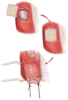
Top: the musculoskeletal defect was created by excising the external and internal oblique layers of the abdominal wall, leaving the transversalus fascia intact. Middle: the test article was implanted in the defect site and secured with Prolene sutures at each of the four corners. Bottom: twenty-six weeks post-implantation, a flap of tissue was created which contained the site of test article placement, identified by the preplaced Prolene sutures. The tissue flap maintained the integrity of the muscular arteries and the thoracic spinal nerve branches that supplied the site of tissue remodeling. The dense connective tissue at the insertion site at the linea alba was connected to the force transducer with silk suture, and positioned such that the direction of the contractile function testing was aligned parallel to the rib origin. Platinum electrodes were placed across the flap proximal and distal to the scaffold placement site.
Figure 2
Figure 2
Tetanic forces of remodeled implants. In-situ muscle contraction studies were performed on muscle flaps from implants at 26 weeks post-surgery and compared to contralateral, native muscle. (A) The tetanic forces (N) generated by remodeled and native tissue following electrical stimulation between 5 and 75 pps. (B) Contractile forces (N/cm2) of tissue, which peaked at 40 pps for the native tissue, and 50 pps for the remodeled tissue. Data expressed as mean ± standard error.
Figure 2
Figure 2
Tetanic forces of remodeled implants. In-situ muscle contraction studies were performed on muscle flaps from implants at 26 weeks post-surgery and compared to contralateral, native muscle. (A) The tetanic forces (N) generated by remodeled and native tissue following electrical stimulation between 5 and 75 pps. (B) Contractile forces (N/cm2) of tissue, which peaked at 40 pps for the native tissue, and 50 pps for the remodeled tissue. Data expressed as mean ± standard error.
Figure 3
Figure 3
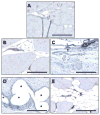
Histologic images of the native muscle tissue and treated remodeled tissue at 26 weeks post surgery. The native tissue (A) was composed mainly of muscle cells organized into well-defined bundles with adjacent mature blood vessels (open arrows) and nerves (closed arrows). The site in which the Restore device was implanted (B) showed new host tissue comprised of bundles of muscle fibers surrounded by vascularized, organized collagenous connective tissue. The site of CuffPatch device implantation (C) showed identifiable remnants of the originally implanted device (asterisks), which were surrounded by dense collagenous tissue, numerous blood vessels, and inflammatory cells. The site in which the polypropylene mesh material was implanted (D) showed highly vascularized, loosely organized connective tissue surrounded the mesh (polymer was displaced from tissue sections due to histologic processing). The site implanted with the excised autologous tissue (E) showed randomly dispersed muscle bundles surrounded by fibrous connective tissue and adipose connective tissue. Tissue sections were stained with Masson's trichrome. Scale bars = 300 μm.
Figure 3
Figure 3
Histologic images of the native muscle tissue and treated remodeled tissue at 26 weeks post surgery. The native tissue (A) was composed mainly of muscle cells organized into well-defined bundles with adjacent mature blood vessels (open arrows) and nerves (closed arrows). The site in which the Restore device was implanted (B) showed new host tissue comprised of bundles of muscle fibers surrounded by vascularized, organized collagenous connective tissue. The site of CuffPatch device implantation (C) showed identifiable remnants of the originally implanted device (asterisks), which were surrounded by dense collagenous tissue, numerous blood vessels, and inflammatory cells. The site in which the polypropylene mesh material was implanted (D) showed highly vascularized, loosely organized connective tissue surrounded the mesh (polymer was displaced from tissue sections due to histologic processing). The site implanted with the excised autologous tissue (E) showed randomly dispersed muscle bundles surrounded by fibrous connective tissue and adipose connective tissue. Tissue sections were stained with Masson's trichrome. Scale bars = 300 μm.
Figure 4
Figure 4
Blood vessels distribution of native and remodeled tissue. Tissue sections at 6 months post surgery were immunolabeled for Von Willibrand Factor to identify endothelial cells associated with the lumen of blood vessels in the native and remodeled tissue sections. Mature blood vessels (A) were located adjacent to the muscle bundles for the native tissue. The tissue repaired with the Restore device (B), and the tissue repaired with the autologous graft (E) showed that larger blood vessels were identified throughout the implantation site. The remnants of the original devices (indicated by *) for CuffPatch (C) and polypropylene mesh (D) were surrounded by a substantial amount of capillaries and larger vessels. Scale bars = 300 μm.
Figure 5
Figure 5
Innervation of native and remodeled tissue. Tissue sections at 6 months post surgery were immunolabeled for anti-neurofilament to identify nerve structures in the native and remodeled tissue sections. Nerves (A) were located adjacent to the muscle bundles for the native tissue. The tissue repaired with the Restore device (B), and the tissue repaired with the autologous graft (E) showed nerves adjacent to the new muscle cells. Individual neurons were located in close proximity to new blood vessels that surrounded the remnants of the CuffPatch device (indicated by *) (C), but no neurons were detected near the polypropylene mesh (indicated by *) (D). Scale bars = 300 μm.
Figure 6
Figure 6
Distribution of slow and fast skeletal muscle fibers in native tissue and remodeled tissue at 26 weeks post surgery. Tissue sections were double-labeled for antimyosin slow (type I) skeletal muscle (brown stain) and antimyosin fast (type II) skeletal muscle (red stain). The native tissue (A) and the tissue repaired with the Restore device (B) showed that the slow muscle fibers were uniformly distributed between the fast muscle fibers. Skeletal muscle fibers surrounded the proximal edges of the CuffPatch device (C) but were not located within the graft site. (D) In the autologous tissue graft, the slow muscle fibers were distributed in an irregular pattern among the fast muscle fibers in the remodeling site, in that the slow fibers were sporadically distributed in some areas and arranged in clusters for other areas. The polypropylene mesh did not show staining for fast or slow skeletal muscle (figure not shown). Scale bars = 300 μm.

References

    1. Akhyari P, Kamiya H, Haverich A, Karck M, Lichtenberg A. Myocardial tissue engineering: the extracellular matrix. Eur J Cardiothorac Surg. 2008;34:229–41. - PubMed
    1. Badylak SF, Kochupura PV, Cohen IS, Doronin SV, Saltman AE, Gilbert TW, et al. The use of extracellular matrix as an inductive scaffold for the partial replacement of functional myocardium. Cell Transplant. 2006;15 1:S29–40. - PubMed
    1. Kochupura PV, Azeloglu EU, Kelly DJ, Doronin SV, Badylak SF, Krukenkamp IB, et al. Tissue-engineered myocardial patch derived from extracellular matrix provides regional mechanical function. Circulation. 2005;112:I144–9. - PubMed
    1. Nieponice A, Gilbert TW, Badylak SF. Reinforcement of esophageal anastomoses with an extracellular matrix scaffold in a canine model. Ann Thorac Surg. 2006;82:2050–8. - PubMed
    1. Nieponice A, McGrath K, Qureshi I, Beckman EJ, Luketich JD, Gilbert TW, et al. An extracellular matrix scaffold for esophageal stricture prevention after circumferential EMR. Gastrointest Endosc. 2008;69:289–96. - PubMed

Publication types

Substances