. 2010 Sep-Oct;80(2-3):99-105.
doi: 10.1016/j.diff.2010.06.007. Epub 2010 Jul 17.

Developmental origin of vaginal epithelium

Affiliations

Developmental origin of vaginal epithelium

Takeshi Kurita. Differentiation. 2010 Sep-Oct.

Abstract

The developmental origin of vaginal epithelium has been controversial for nearly a century, with speculation that vaginal epithelium originates from the Müllerian duct, Wolffian duct, and/or urogenital sinus. None of these possibilities have been definitively proven or disproven by direct scientific data. To define precisely the origin of vaginal epithelium, epithelial cells of the Müllerian duct, Wolffian duct, or urogenital sinus were fluorescently labeled in mouse embryos by crossing tdTomato-EGFP dual-reporter transgenic mice with transgenic mouse lines that express Cre-recombinase in each type of epithelium. In embryos and newborn mice, the vagina consisted of fused Müllerian ducts plus the sinus vagina of urogenital sinus origin. However, the proportion of the sinus vagina was significantly reduced as the Müllerian vagina grew caudally. By postpartum day 7, the Müllerian vagina extended to the caudal end of the body, whereas the sinus vagina remained only at the junction between the vagina and perineal skin. As the vagina opened in puberty, urogenital sinus epithelium was detected only in the vulva, but not in the vagina. Additionally, from embryo to adult stages, residual Wolffian duct epithelium was present in the dorsolateral stromal wall of the vagina, but not within vaginal or vulvar epithelium. In conclusion, adult mouse vaginal epithelium is derived solely from Müllerian duct epithelium.


Figures

Figure 1
Figure 1
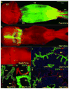
Expression of EGFP in embryonic MDE, WDE, and UGE. Phase-contrast (a-c), phase-contrast + fluorescence (d-f) images are shown. The Müllerian vagina (MV), vaginal bulb (VB), and WDs (indicated by *) were present in the E17 vagina (a-c). EGFP signal identifies epithelial cells originating from the WDs and MDs in Pax2-Cre (d), WDs in Hoxb7-Cre (e), and UGS in Osr1-Cre (f) reporter mice. The dotted lines indicate the outline of the VB. While the entire VB epithelium was positive for EGFP in Osr1-Cre reporter mouse embryos (f), EGFP was absent in the VB of Pax2-Cre and Hoxb7-Cre reporter mouse embryos, indicating that the VB arises from the UGS.
Figure 2
Figure 2
Cellular origin of neonatal vaginal epithelium. EGFP expression was analyzed in female Pax2-Cre (expressed in MDE+WDE, a), Osr1-Cre (expressed in UGE, b, d, e, g and h), and Hoxb7-Cre (expressed in WDE, c and f) reporter mice on P0 (a-c) and P3 (d-h). Longitudinal (e) and transverse (f-h) sections of the female genital tract were stained with DAPI (f-h; blue) and the expression of EGFP (green) and p63 (red) in Hoxb7-Cre reporter mice (f) or Pax2 (green) and p63 (red) in Osr1-Cre reporter mice (g-h) were analyzed by dual-immunofluorescence. On P0, the vagina consisted of Müllerian vagina (MV, a) and sinus vagina (SV, b). Fragments of WDE were also present at the junction of MV and SV (c, indicated by *). On P3, EGFP was detected only in the solid epithelium but not in the canalized epithelium in Osr1-Cre reporter mice (d, e). Thus, the vaginal canal in the lower vagina formed by caudal growth of the MV. Also, UGE in the SV (in Osr1-Cre reporter mice) was found only at the caudal end of vagina (the junction between the SV and MV is indicated by the arrows in d and e). The epithelia in the SV and urethra (UR; positive for EGFP in Osr1-Cre reporter mice) were still connected within external genitalia (EG, d). Transverse images (f-h) represent P3 vaginal tissue sections from caudal (f, connected with urethra) to crainial (h, junction between SV and MV). The solid epithelium of the SV formed a plate-like cord (g, h, p63-positive and Pax2-negative). In contrast, epithelium in the MV formed a flat, tube shape (h, Pax2- and p63-positive [in the basal layer]; the dual-positive cells for pax2 and p63 appear yellow). The WDE (EGFP-positive in Hoxb7-Cre reporter mice, indicated by *) was negative for p63. The WDE was occasionally detected in association with the SV at its caudal end (f, Intrusion of a single WD epithelial cell into the SV is indicated by the arrow). In other sections of the vagina, the WDE was located within the stroma separated from vaginal epithelium (g, Pax2-positive and p63 negative).
Figure 3
Figure 3
Distribution of MDE, WDE, and UGE in the lower reproductive tract of the female neonatal mouse. Expression of EGFP was analyzed in female Pax2-Cre (a, d), Osr1-Cre (b, eg), and Hoxb7-Cre (c) reporter mice at P8 (a-c) and P12 (d-g). In Pax2-Cre reporter mice, the entire epithelium in the vagina (VG), uterus (UT), and cervix (CVX) was positive for EGFP, indicating its MDE origin (a). Solid epithelium of UGS origin was present at the junction between vagina and external genitalia (EG) (b). Streaks/fragments of residual WDE were still present in the dorsal wall of the lower vagina in Pax2-Cre and Hoxb7-Cre reporter mice (a, c, indicated by *). Expression of EGFP, Pax2, and p63 at the junction of the Müllerian vagina (MV) and sinus vagina (SV) was assessed by dual-immunofluorescence of transverse sections (d-g, dorsal side up, bar = 50 μm). As observed in earlier stages, the vaginal canal was lined with MDE, which was EGFP-positive in Pax2-Cre reporter mice (d) but EGFP-negative in Osr1-Cre reporter mice (e-g). Throughout the development of the vagina, Pax2 was expressed in epithelial cells of the MDs but was of UGS origin; therefore, the vaginal canal was lined with Pax2-positive epithelium of MD origin (e).
Figure 4
Figure 4
Distribution of MDE, WDE, and UGE in the vagina and vulva at puberty. The distribution of EGFP-positive cells was examined in the vaginal openings of 4-week-old female Pax2-Cre (a) and Osr1-Cre (b) reporter mice. MDE and UGE were distributed in a complementary pattern; MDE was present in the vagina (vg) but not the vulva (vu), whereas UGE was present in the vulva but not the vagina. The presence of EGFP-positive cells in vaginal epithelium was examined through the surface of the entire vagina in Pax2-Cre (c), Hoxb7-Cre (not shown), and Osr1-Cre (d) reporter mice. Caudal is to the left in panels c and d. EGFP-positive cells were present in the vaginal epithelium of Pax2-Cre, but not Osr1-Cre or Hoxb7-Cre, reporter mice. Bars = 0.5 mm.

Comment in

References

    1. Arey LB. Developmental anatomy: a textbook and laboratory manual of embryology. W.B. Saunders; Philadelphia: 1954.
    1. Bloomfield A, Frazer JE. The Development of the Lower End of the Vagina. Journal of anatomy. 1927;62:9–32. - PMC - PubMed
    1. Boutin E, Sanderson R, Bernfield M, Cunha GR. Expression of syndecan, a cell surface proteoglycan, correlates with induced changes in cellular organization. The Journal of cell biology. 1989;107:605a.
    1. Boutin EL, Sanderson RD, Bernfield M, Cunha GR. Epithelial-mesenchymal interactions in uterus and vagina alter the expression of the cell surface proteoglycan, syndecan. Developmental biology. 1991;148:63–74. - PubMed
    1. Branda CS, Dymecki SM. Talking about a revolution: The impact of site-specific recombinases on genetic analyses in mice. Developmental cell. 2004;6:7–28. - PubMed

Publication types