. 2010 Sep;31(27):6941-6951.
doi: 10.1016/j.biomaterials.2010.05.056. Epub 2010 Jun 19.

Directed 3D cell alignment and elongation in microengineered hydrogels

Affiliations

Directed 3D cell alignment and elongation in microengineered hydrogels

Hug Aubin et al. Biomaterials. 2010 Sep.

Abstract

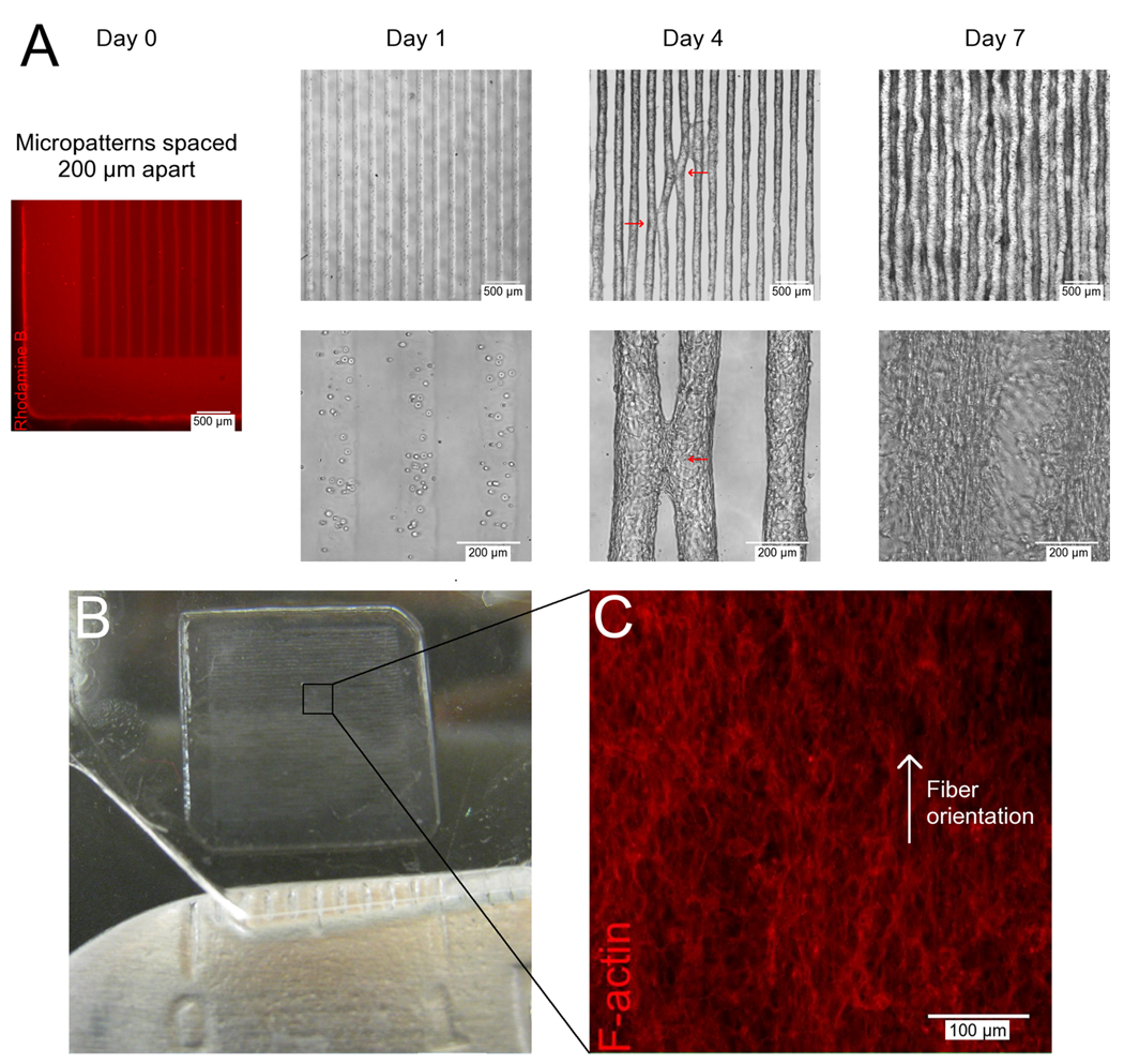
Organized cellular alignment is critical to controlling tissue microarchitecture and biological function. Although a multitude of techniques have been described to control cellular alignment in 2D, recapitulating the cellular alignment of highly organized native tissues in 3D engineered tissues remains a challenge. While cellular alignment in engineered tissues can be induced through the use of external physical stimuli, there are few simple techniques for microscale control of cell behavior that are largely cell-driven. In this study we present a simple and direct method to control the alignment and elongation of fibroblasts, myoblasts, endothelial cells and cardiac stem cells encapsulated in microengineered 3D gelatin methacrylate (GelMA) hydrogels, demonstrating that cells with the intrinsic potential to form aligned tissues in vivo will self-organize into functional tissues in vitro if confined in the appropriate 3D microarchitecture. The presented system may be used as an in vitro model for investigating cell and tissue morphogenesis in 3D, as well as for creating tissue constructs with microscale control of 3D cellular alignment and elongation, that could have great potential for the engineering of functional tissues with aligned cells and anisotropic function.


Figures

Figure 1
Figure 1
Cell morphology and organization as a function of time in patterned and unpatterned microconstructs. Patterning 3T3-fibroblast-laden 5% GelMA hydrogels into high aspect ratio rectangular microconstructs (50 µm (w) × 800 µm (l) × 150 µm (h)) induced cellular alignment and elongation, while cellular orientation in unpatterned hydrogels remained random. (A) Rhodamine B stained GelMA hydrogel construct shows the patterned and unpatterned regions. (B) Representative phase contrast images of 3T3-fibroblasts (10 × 106 cells/mL) encapsulated in patterned (top row) and unpatterned regions of the GelMA hydrogel (bottom row) on days 1, 3 and 5 of culture show that elongation increases over time for all hydrogels, while alignment is only induced in patterned constructs.
Figure 2
Figure 2
Multilayer analysis of cell alignment. Cellular alignment in 3T3-fibroblast-laden 5% GelMA hydrogels patterned into high aspect ratio rectangular microconstructs (50 µm (w) × 800 µm (l) × 150 µm (h)) was maintained throughout the entire height of the construct. (A) Representative DAPI/F-actin stained 3D-projection (50 µm z-stack) of a 3T3-laden rectangular microconstruct after 5 days of culture. (B) Three DAPI and F-actin stained xy-planes from the microconstruct (distance: 10 µm in z-plane) with corresponding histograms ((C), (D) and (E) respectively) showing the nuclear alignment in 10° increments relative to the preferred nuclear orientation of each individual plane demonstrates similar alignment along the major axis throughout the thickness of the constructs.
Figure 3
Figure 3
Cell elongation and alignment as a function of microconstruct width. Decreasing the width of patterned rectangular 5% GelMA microstructures (800 µm (l) × 150 µm (h)) increased 3T3-fibroblast alignment as well as nuclear elongation after 5 days of culture. (A) Mean percentage of aligned cell nuclei (within 10° of preferred nuclear orientation) shows 100 µm constructs were significantly more aligned than unpatterned controls, while 50 µm constructs were significantly more aligned than all other groups. (B) Mean nuclear shape index (circularity = 4*π*area/perimeter2) shows a similar pattern with 100 µm constructs having a significantly lower index than unpatterned controls, while 50 µm constructs were significantly different than all other groups. (C), (D), (E) and (F) Histograms of the relative alignment in 10° increments demonstrates increased cellular alignment with decreased microconstruct width. (G), (H), (I) and (J) Representative phase contrast images of unpatterned, 200 µm, 100 µm and 50 µm wide microconstructs respectively show significantly increased cell alignment and elongation inside the constructs with decreasing width. (Error bars: ± SD; *** p<0.001; ** p<0.01; * p<0.01)
Figure 4
Figure 4
Effect of MMP inhibition on elongation and alignment in patterned microconstructs. General MMP inhibition with doxycycline supplemented media (400 µM) for 4 days of culture decreased nuclear alignment and elongation in 3T3-fibroblast-laden 5% GelMA hydrogels patterned into high aspect ratio rectangular microconstructs (50 µm (w) × 800 µm (l) × 150 µm (h)) and showed no differences to unpatterned hydrogels with and without MMP inhibition. (A) Zymogram showing MMP-2 and MMP-9 activity in culture media of cell-laden micropatterned hydrogels compared to human standard. (B) Normalized MMP-2 and MMP-9 activity of unsupplemented and supplemented samples demonstrates significant decreases in MMP expression and activity with inhibition. (C) Mean cell nuclei alignment (within 10° of preferred nuclear orientation) shows decreased alignment with MMP inhibition. (D) Mean nuclear shape index (circularity = 4*π*area/perimeter2) similarly shows decreased cell elongation with MMP inhibition, however micropatterned constructs show significantly greater elongation than unpatterned controls. (E) and (F) Histograms showing the percentage of aligned cell nuclei in 10° increments, with representative z-stack overlays of DAPI/F-actin staining of microconstructs cultured in unsupplemented and supplemented media respectively. (Error bars: ± SD; *** p<0.001; * p<0.01)
Figure 5
Figure 5
Control of cell elongation and alignment in multiple cell types. Patterning cell-laden 5% GelMA hydrogels into rectangular microconstructs (50 µm (w) × 800 µm (l) × 150 µm (h)) induced cellular alignment and elongation after 5 days of culture only in cell types possessing an intrinsic potential to organize into aligned tissues in vivo, while cellular orientation in unpatterned parts of the same hydrogels remained random. Representative z-stack overlays of DAPI/F-actin staining of (A) patterned and (B) unpatterned hydrogels laden with HUVEC, C2C12, CSP, and Hep-G2 cells respectively show patterning induced aligned and elongated cell-network formations in HUVEC, C2C12 and CSP cells and patterning independent cell-cluster formations in Hep-G2 cells. (C) Mean percentage of aligned cell nuclei (within 10° of preferred nuclear orientation) shows that patterned HUVEC-, C2C12- and CSP-laden constructs were significantly more aligned than unpatterned controls, while in Hep-G2-laden constructs patterning failed to induce cell alignment. (D) Mean nuclear shape index (circularity = 4*π*area/perimeter2) similarly shows that patterned HUVEC-, C2C12- and CSP-laden constructs were significantly more elongated than unpatterned controls, while in Hep-G2-laden constructs patterning failed to induce cell elongation. (Error bars: ± SD; ** p< 0.01; *** p<0.001)
Figure 6
Figure 6
Microconstruct width as a function of time in patterned cell-laden hydrogels. 3T3-fibroblast-laden 5% GelMA hydrogels patterned into rectangular microconstructs (50 µm (w)×800 µm (l)×150 µm (h)) increased in width after initial swelling due to cell proliferation. (A) Mean microconstruct width at each culture day after initial 24 h swelling period shows increase in feature size. (B) Rhodamine B stained hydrogel construct demonstrates the initial micropattern width of 50 µm directly after patterning. (C) and (D) Representative phase contrast images of a single microconstruct at day 1 and 5 respectively show increase in feature size due to cell proliferation. (Error bars: ± SD; *** p< 0.001)
Figure 7
Figure 7
Self-assembly of multiple aligned microconstructs into a macroscale and aligned 3D tissue construct. 3T3-fibroblast-laden 5% GelMA hydrogels patterned into rectangular microconstructs (50 µm (w) × 800 µm (l) × 150 µm (h)) spaced 200 µm apart self-assembled into macroscale and aligned 3D tissue constructs after 7 days of culture through convergence of multiple, aligned microconstructs. (A) Rhodamine B stained hydrogel shows initial microconstruct spacing of 200 µm at day 0; representative phase contrast images of cell-laden microconstructs at day 1, 4 and 7 respectively show focal points of contact between neighboring aligned microconstructs at day 4 (red arrows) and convergence into a macroscale 3D tissue construct at day 7. (B) Image of a 1 cm × 1 cm, self-assembled 3D tissue construct at day 7. (C) Representative F-Actin staining of middle xy-plane of macroscale 3D tissue construct shows orientated actin fiber organization in single direction.

References

    1. Papadaki M, Bursac N, Langer R, Merok J, Vunjak-Novakovic G, Freed LE. Tissue engineering of functional cardiac muscle: Molecular, structural, and electrophysiological studies. Am J Physiol Heart Circ Physiol. 2001;280(1):H168–H178. - PubMed
    1. Wigmore PM, Dunglison GF. The generation of fiber diversity during myogenesis. Int J Dev Biol. 1998;42(2):117–125. - PubMed
    1. Chiu JJ, Chen LJ, Chen CN, Lee PL, Lee CI. A model for studying the effect of shear stress on interactions between vascular endothelial cells and smooth muscle cells. J Biomech. 2004;37(4):531–539. - PubMed
    1. Vunjak-Novakovic G, Altman G, Horan R, Kaplan DL. Tissue engineering of ligaments. Annu Rev Biomed Eng. 2004:6131–6156. - PubMed
    1. Khademhosseini A, Vacanti JP, Langer R. Progress in tissue engineering. Sci Am. 2009;300(5):64–71. - PubMed