. 2010 Sep;192(18):4562-70.
doi: 10.1128/JB.00536-10. Epub 2010 Jul 16.

Massive gene duplication event among clinical isolates of the Mycobacterium tuberculosis W/Beijing family

Affiliations

Massive gene duplication event among clinical isolates of the Mycobacterium tuberculosis W/Beijing family

Pilar Domenech et al. J Bacteriol. 2010 Sep.

Abstract

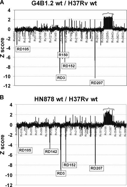
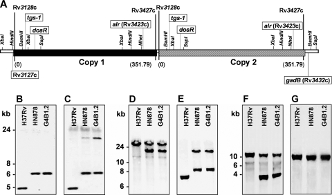
As part of our effort to uncover the molecular basis for the phenotypic variation among clinical Mycobacterium tuberculosis isolates, we have previously reported that isolates belonging to the W/Beijing lineage constitutively overexpress the DosR-regulated transcriptional program. While generating dosR knockouts in two independent W/Beijing sublineages, we were surprised to discover that they possess two copies of dosR. This dosR amplification is part of a massive genomic duplication spanning 350 kb and encompassing >300 genes. In total, this equates to 8% of the genome being present as two copies. The presence of IS6110 elements at both ends of the region of duplication, and in the novel junction region, suggests that it arose through unequal homologous recombination of sister chromatids at the IS6110 sequences. Analysis of isolates representing the major M. tuberculosis lineages has revealed that the 350-kb duplication is restricted to the most recently evolved sublineages of the W/Beijing family. Within these isolates, the duplication is partly responsible for the constitutive dosR overexpression phenotype. Although the nature of the selection event giving rise to the duplication remains unresolved, its evolution is almost certainly the result of specific selective pressure(s) encountered inside the host. A preliminary in vitro screen has failed to reveal a role of the duplication in conferring resistance to common antitubercular drugs, a trait frequently associated with W/Beijing isolates. Nevertheless, this first description of a genetic remodeling event of this nature for M. tuberculosis further highlights the potential for the evolution of diversity in this important global pathogen.


Figures

FIG. 1.
FIG. 1.
Southern blotting reveals the presence of two copies of dosR in recombinant W/Beijing strains. Genomic DNA from wild-type and two independent dosR::hyg recombinants generated for H37Rv (clones 19 and 24) and the W/Beijing isolates HN878 (clones 25 and 28) and G4B1.2 (clones 30 and 32) was digested with PstI and transferred by Southern blotting. (A) The membrane was hybridized with a 2.4-kb PCR fragment (generated using dosR-1 and dosS-1) that includes Rv3134c, dosR, and 739 bp of dosS. Note the presence of an intact copy of dosR retained by the HN878 and G4B1.2 dosR::hyg strains. (B) Based on the published H37Rv sequence, a genetic map of the dosR region in the recombinant strains is shown. By replacing 486 bp of dosR with the hyg resistance cassette, an additional PstI site has been introduced.
FIG. 2.
FIG. 2.
Characterization of the W/Beijing duplication through whole-genome microarray. Comparative array hybridizations were carried out for genomic DNA prepared from wild-type W/Beijing isolates G4B1.2 (A) and HN878 (B) versus H37Rv. For each, the average Z scores from three arrays for each of the 3,924 genes present in H37Rv are shown. The RD105 and RD207 deletions unique to the W/Beijing lineage are indicated, as are the RD150 and RD142 deletions that are characteristic of group 4 and 5 W/Beijing isolates, respectively. The RD3 and RD152 deletions were also identified. The majority of the ∼300 genes in the contiguous stretch from Rv3128c to Rv3427c showed a Z score of ≥2.0, consistent with the presence of a 350-kb duplicated region in G4B1.2 and HN878 relative to H37Rv.
FIG. 3.
FIG. 3.
The two copies of the 350-kb duplicated region are arranged in tandem. (A) A schematic representation of the duplicated region is shown. Genes Rv3128c and Rv3427c mark the approximate beginning and end of each duplicated segment, respectively. Genes Rv3127c and gadB flank the duplication externally. The length of the duplication is indicated in parentheses, and the two copies (Copy 1 and Copy 2) are arranged in a direct, tandem duplication. The arrangement of the genes indicated (boxed) was confirmed by Southern blotting of genomic DNA comparing wild-type H37Rv and the W/Beijing isolates HN878 and G4B1.2. The approximate locations of the restriction enzyme sites are based upon the published H37Rv sequence. (B and C) Southern blots confirming that the beginning of the duplication is located between Rv3127c (single copy) and tgs1 (duplicated). DNAs were digested with XbaI and hybridized with a 620-bp probe derived from Rv3127c (B; primers Rv3127c-A and -B) or with a 920-bp tgs1 probe (C; primers Rv3130-1 and Rv3130c-F). (D and E) Southern blots confirming the location of the junction of the duplication and that the two copies are arranged in a direct, tandem duplication. DNAs digested with HindIII and SspI were hybridized with a 540-bp alr probe (D; primers alr-C and alr-R) or with a 1-kb dosR probe [E; primers dosR-C and dosRrev(HindIII)]. (F and G) Southern blots confirming that the end of the duplication is located between Rv3427c (duplicated) and gadB (single copy). DNAs were digested with BamHI and NheI and hybridized with the 540-bp alr probe (F) or a 1.2-kb gadB probe (G; primers gadB-C and gadB-D).
FIG. 4.
FIG. 4.
Genetic map and PCR-based screening for identifying isolates bearing the 350-kb duplication. The genetic arrangement of the cloned fragments representing the beginning (A), end (B), and junction (C) regions of the duplication is shown. Genes present in “Copy 1” of the duplication are indicated in black, while those in “Copy 2” are in dark gray. The three sets of primers used to screen for the presence of the duplication in clinical isolates are highlighted (boxed). The screening method is based on detecting the IS6110 transposase insertions (white) located in the beginning (A and D), end (B and E), and junction (C and F) regions. (D to F) Lanes: 1, M. tuberculosis West African-I; 2, M. bovis BCG; 3, M. bovis; 4, M. canetti; 5 and 6, M. tuberculosis Indo-Oceanic (I-O) lineage; 7 and 8, East African-Indian (E-A-I); 9 and 10, Euro-American (E-A); 11 and 12, W/Beijing group 1 (G-1); 13 and 14, W/Beijing group 2 (G-2); 15 to 20, W/Beijing group 3 (G-3); 21 to 24, W/Beijing group 4 (G-4); 25 to 28, W/Beijing group 5 (G-5); 29, negative control. In the example shown, the W/Beijing isolates in lanes 23 and 24 (group 4) and in lanes 27 and 28 (group 5) are positive in all three reactions, suggesting that they contain the full duplication.
FIG. 5.
FIG. 5.
Impact of the W/Beijing duplication on dosR expression. (A) qRT-PCR analysis of dosR expression in the wild type (wt) and the single or double dosR mutant strains indicated in panel B. The expression levels of dosR are normalized to the sigA housekeeping gene and are plotted relative to H37Rv. Each sample was assayed in quadruplicate, and at least two independent biological replicates were analyzed for each strain. Data from a single representative experiment are presented. Error bars represent standard deviation. (B) Schematic representation depicting the duplication and dosR genotypes for each of the strains analyzed in panel A. Further details for each strain can be found in the text.

References

    1. Alland, D., D. W. Lacher, M. H. Hazbon, A. S. Motiwala, W. Qi, R. D. Fleischmann, and T. S. Whittam. 2007. Role of large sequence polymorphisms (LSPs) in generating genomic diversity among clinical isolates of Mycobacterium tuberculosis and the utility of LSPs in phylogenetic analysis. J. Clin. Microbiol. 45:39-46. - PMC - PubMed
    1. Andersson, D. I., and D. Hughes. 2009. Gene amplification and adaptive evolution in bacteria. Annu. Rev. Genet. 43:167-195. - PubMed
    1. Bifani, P. J., B. Mathema, N. E. Kurepina, and B. N. Kreiswirth. 2002. Global dissemination of the Mycobacterium tuberculosis W-Beijing family strains. Trends Microbiol. 10:45-52. - PubMed
    1. Brosch, R., S. V. Gordon, T. Garnier, K. Eiglmeier, W. Frigui, P. Valenti, S. Dos Santos, S. Duthoy, C. Lacroix, C. Garcia-Pelayo, J. K. Inwald, P. Golby, J. N. Garcia, R. G. Hewinson, M. A. Behr, M. A. Quail, C. Churcher, B. G. Barrell, J. Parkhill, and S. T. Cole. 2007. Genome plasticity of BCG and impact on vaccine efficacy. Proc. Natl. Acad. Sci. U. S. A. 104:5596-5601. - PMC - PubMed
    1. Brosch, R., S. V. Gordon, M. Marmiesse, P. Brodin, C. Buchrieser, K. Eiglmeier, T. Garnier, C. Gutierrez, G. Hewinson, K. Kremer, L. M. Parsons, A. S. Pym, S. Samper, D. van Soolingen, and S. T. Cole. 2002. A new evolutionary scenario for the Mycobacterium tuberculosis complex. Proc. Natl. Acad. Sci. U. S. A. 99:3684-3689. - PMC - PubMed

Publication types

MeSH terms

Associated data