. 2010 Aug 15;185(4):2200-8.
doi: 10.4049/jimmunol.1000332. Epub 2010 Jul 16.

In hepatic fibrosis, liver sinusoidal endothelial cells acquire enhanced immunogenicity

Affiliations

In hepatic fibrosis, liver sinusoidal endothelial cells acquire enhanced immunogenicity

Michael K Connolly et al. J Immunol. .

Abstract

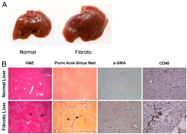
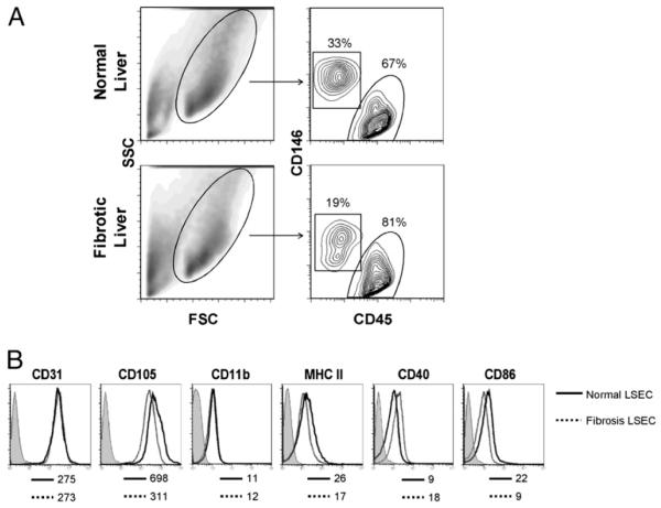
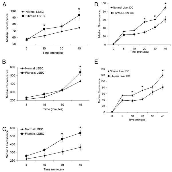
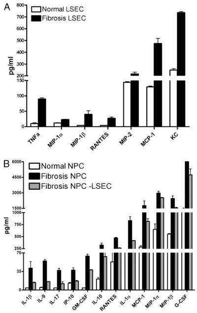
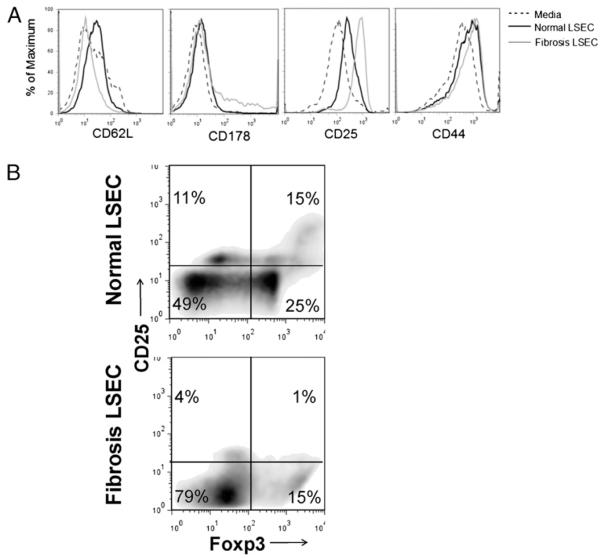
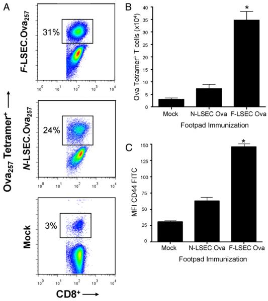
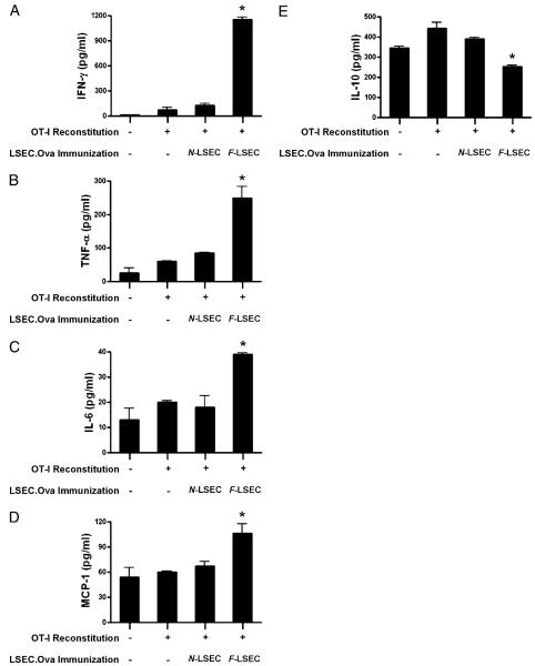
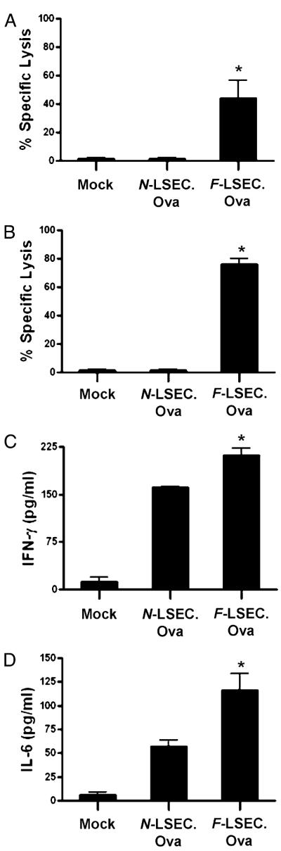
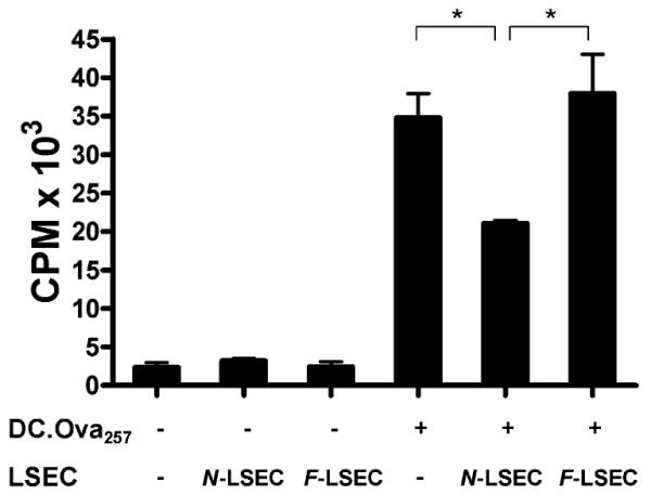
The normal liver is characterized by immunologic tolerance. Primary mediators of hepatic immune tolerance are liver sinusoidal endothelial cells (LSECs). LSECs block adaptive immunogenic responses to Ag and induce the generation of T regulatory cells. Hepatic fibrosis is characterized by both intense intrahepatic inflammation and altered hepatic immunity. We postulated that, in liver fibrosis, a reversal of LSEC function from tolerogenic to proinflammatory and immunogenic may contribute to both the heightened inflammatory milieu and altered intrahepatic immunity. We found that, after fibrotic liver injury from hepatotoxins, LSECs become highly proinflammatory and secrete an array of cytokines and chemokines. In addition, LSECs gain enhanced capacity to capture Ag and induce T cell proliferation. Similarly, unlike LSECs in normal livers, in fibrosis, LSECs do not veto dendritic cell priming of T cells. Furthermore, whereas in normal livers, LSECs are active in the generation of T regulatory cells, in hepatic fibrosis LSECs induce an immunogenic T cell phenotype capable of enhancing endogenous CTLs and generating potent de novo CTL responses. Moreover, depletion of LSECs from fibrotic liver cultures mitigates the proinflammatory milieu characteristic of hepatic fibrosis. Our findings offer a critical understanding of the role of LSECs in modulating intrahepatic immunity and inflammation in fibro-inflammatory liver disease.


Figures

FIGURE 1
FIGURE 1
Model of liver fibrosis. A, The liver surface contour of thioacetamide/leptin-treated mice at 8 wk is markedly irregular and exhibits diffuse nodularity. B, H&E examination (original magnification×10) of the livers of treated mice reveals a distorted hepatic architectural pattern. Regenerative nodules are seen (asterisk) bounded by fibrous septa (arrows). Picric acid-Sirius red (original magnification×10) staining highlights the collagen network both surrounding and bridging the portal triads. α-SMA staining (original magnification×10) is prominent in the livers of treated mice, suggesting HSC activation. CD45 (original magnification×20) immunohistochemistry confirms the presence of a lymphocytic infiltrate in the livers of treated animals.
FIGURE 2
FIGURE 2
LSEC surface phenotype in fibrosis. A, Live NPCs from normal and fibrotic livers were analyzed for expression of both CD45 and CD146. Dead cells were excluded using 7-aminoactinomycin D (data not shown). B, CD45CD146+ LSECs were gated and further analyzed for cell surface marker expression. Isotype staining, shown in shaded histograms for N-LSEC, was similar for both groups. Mean fluorescence index is indicated for each marker below their respective histograms. Data are representative of experiments repeated more than three times using three to four mice per experiment. FSC, forward light scatter; SSC, side light scatter.
FIGURE 3
FIGURE 3
Ag capture by LSECs is altered in hepatic fibrosis. LSECs (5 × 10⁵) were tested for their ability to capture dextran (A), albumin (B), and mannose-albumin (C) in vitro at various time points of incubation. Similarly, DCs (5 × 10⁵) from normal and fibrotic livers were tested for their ability to capture albumin (D) and mannose-albumin (E). Experiments were repeated more than three times and performed in triplicate. *p < 0.05.
FIGURE 4
FIGURE 4
LESC are proinflammatory in hepatic fibrosis. A, Freshly isolated LSECs were cultured for 24 h, and inflammatory mediators were measured in cell culture supernatant. p < 0.05 for each comparison. B, NPCs were harvested from the livers of normal and fibrotic mice and cultured for 24 h. Depletion of LSECs from fibrotic NPC concentrates by FACS abrogated their elevated cytokine and chemokine production. Experiments were repeated more than three times and performed in triplicate. p < 0.05 for each inflammatory mediator.
FIGURE 5
FIGURE 5
LSECs from fibrotic livers are immunogenic. Freshly isolated LSECs (2 × 10⁵) from normal or fibrotic livers were either A, loaded with OVA257–264 and cocultured with CD8+ OT-I T cells (2 × 10⁵) before measurement of T cell activation markers or B, coincubated with CD4+ T cells (2 × 10⁵) from C57BL/6 mice before determination of CD25 and Foxp3 coexpression. Data are representative of experiments repeated three times.
FIGURE 6
FIGURE 6
F-LSECs produce enhanced T cell proliferation in vivo. Mice were adoptively transferred with OT-I T cells (2 × 10⁶) before receiving footpad immunization with LSEC. OVA257 (2 × 10⁵) or mock immunized. At 96 h, ipsilateral popliteal lymph nodes were harvested and costained for CD8 and OVA tetramer. The fraction of OVA tetramer+ cells among all CD8+ cells (A), the total number of Ova tetramer+ cells (B), and their expression of CD44 (C) were determined by flow cytometry. Experiments were repeated three times using two to four mice per experiment. *p < 0.05.
FIGURE 7
FIGURE 7
F-LSECs enhance endogenous CTL. Rag mice were reconstituted with OT-I T cells (1 × 10⁷) before either mock immunization or immunization using LSEC.OVA257 (1 × 10⁶). One week postimmunization, splenocytes from treated animals were restimulated in vitro with OVA257–264 before measurement of IFN-γ (A), TNF-α (B), IL-6 (C), MCP-1 (D), and IL-10 (E) in day 5 CTL cultures. Experiments were repeated twice using two to four mice per experiment. *p < 0.05.
FIGURE 8
FIGURE 8
F-LSECs induce potent de novo CTL responses. Naive C57BL/6 mice were either mock immunized or twice immunized using LSEC.OVA257 (1 × 10⁶). One week later, in vivo CTL lysis of OVA257-presenting cellular targets was measured in the spleen (A) and the liver (B), and splenocyte CTL cultures were assayed for production of IFN-γ (C) and IL-6 (D). Experiments were repeated three times and performed in triplicate using two to four mice per group. *p < 0.05.
FIGURE 9
FIGURE 9
F-LSECs do not mitigate Ag presentation by DC. OT-I T cells (1 × 10⁵) were cultured alone or coincubated with DC.OVA257–264 (1 × 10⁴) and LSECs (1 × 10⁴), separately or in combination, harvested from normal or fibrotic livers. DC.OVA257 induced potent Ag-specific T cell proliferation that was mitigated by coculture with N-LSECs. Conversely, coculture with F-LSECs did not lower DC-mediated T cell proliferation. Experiments were repeated three times and performed in triplicate. *p < 0.05.

References

    1. Elvevold K, Smedsrød B, Martinez I. The liver sinusoidal endothelial cell: a cell type of controversial and confusing identity. Am. J. Physiol. Gastrointest. Liver Physiol. 2008;294:G391–G400. - PubMed
    1. Braet F, Wisse E. Structural and functional aspects of liver sinusoidal endothelial cell fenestrae: a review. Comp. Hepatol. 2002;1:1. - PMC - PubMed
    1. Katz SC, Pillarisetty VG, Bleier JI, Shah AB, DeMatteo RP. Liver sinusoidal endothelial cells are insufficient to activate T cells. J. Immunol. 2004;173:230–235. - PubMed
    1. Schurich A, Böttcher JP, Burgdorf S, Penzler P, Hegenbarth S, Kern M, Dolf A, Endl E, Schultze J, Wiertz E, et al. Distinct kinetics and dynamics of cross-presentation in liver sinusoidal endothelial cells compared to dendritic cells. Hepatology. 2009;50:909–919. - PubMed
    1. Knolle PA, Limmer A. Neighborhood politics: the immunoregulatory function of organ-resident liver endothelial cells. Trends Immunol. 2001;22:432–437. - PubMed

Publication types

MeSH terms