. 2010 Aug 18;29(16):2802-12.
doi: 10.1038/emboj.2010.157. Epub 2010 Jul 16.

14-3-3gamma mediates Cdc25A proteolysis to block premature mitotic entry after DNA damage

Affiliations

14-3-3gamma mediates Cdc25A proteolysis to block premature mitotic entry after DNA damage

Kousuke Kasahara et al. EMBO J. .

Abstract

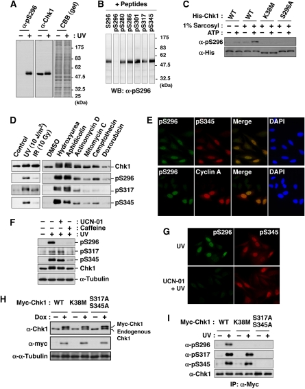
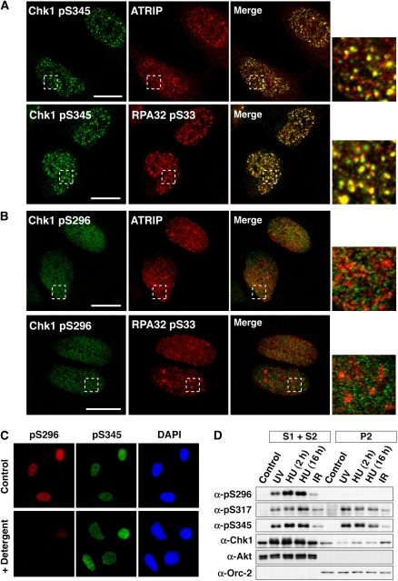
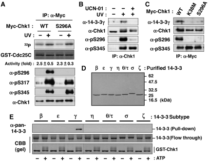
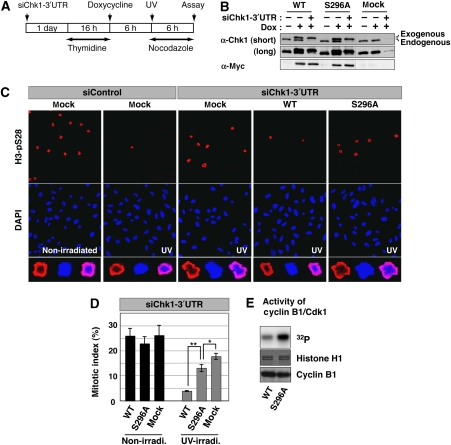
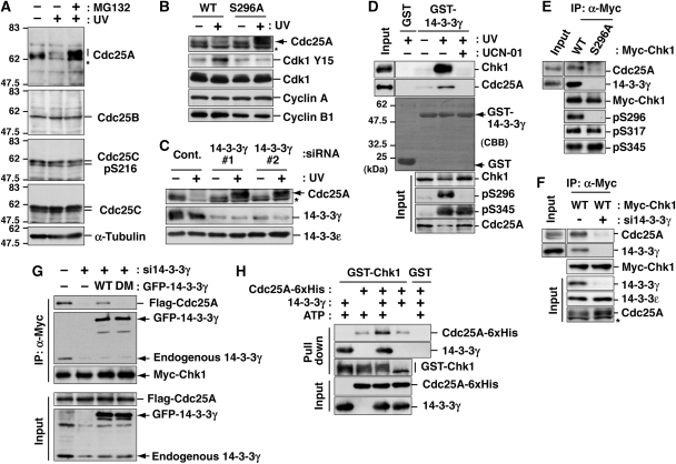
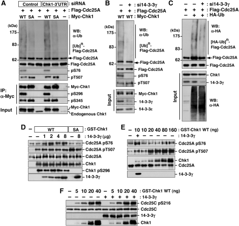
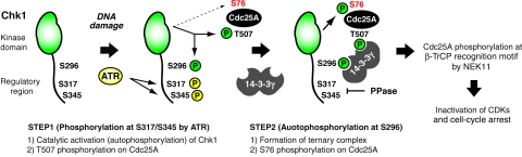
14-3-3 proteins control various cellular processes, including cell cycle progression and DNA damage checkpoint. At the DNA damage checkpoint, some subtypes of 14-3-3 (beta and zeta isoforms in mammalian cells and Rad24 in fission yeast) bind to Ser345-phosphorylated Chk1 and promote its nuclear retention. Here, we report that 14-3-3gamma forms a complex with Chk1 phosphorylated at Ser296, but not at ATR sites (Ser317 and Ser345). Ser296 phosphorylation is catalysed by Chk1 itself after Chk1 phosphorylation by ATR, and then ATR sites are rapidly dephosphorylated on Ser296-phosphorylated Chk1. Although Ser345 phosphorylation is observed at nuclear DNA damage foci, it occurs more diffusely in the nucleus. The replacement of endogenous Chk1 with Chk1 mutated at Ser296 to Ala induces premature mitotic entry after ultraviolet irradiation, suggesting the importance of Ser296 phosphorylation in the DNA damage response. Although Ser296 phosphorylation induces the only marginal change in Chk1 catalytic activity, 14-3-3gamma mediates the interaction between Chk1 and Cdc25A. This ternary complex formation has an essential function in Cdc25A phosphorylation and degradation to block premature mitotic entry after DNA damage.


Conflict of interest statement

The authors declare that they have no conflict of interest.

Figures

Figure 1
Figure 1
Chk1 autophosphorylation at Ser296. (A, B) Characterization of an antibody specifically recognizing Chk1 phosphorylation at Ser296. The antibody (α-pS296) reacted specifically with a band corresponding to Chk1 in lysates of UV-irradiated (+) HeLa cells (Ikegami et al, 2008), but not of non-treated (−) cells (A). Immunoreactivity was impaired specifically by preincubation with pS296 corresponding to Ser296-phosphorylated Chk1, but not with non-phosphorylated peptide S296 and phosphopeptides for other sites within Chk1 (B). (C) The in vitro autophosphorylation assay using 6xHis-ProS2-Chk1 (His-Chk1) expressed in bacteria. E. coli strain BL21-CodonPlus®(DE3)-RP was transformed with pCold ProS2 carrying each Chk1. Each 6xHis-ProS2-tagged Chk1 was expressed in the presence of 1 mM IPTG at 15°C for 16 h. E. coli cells were lysed in the lysis buffer (40 mM Hepes-NaOH [pH 8.0], 300 mM NaCl, 1% Triton X-100) supplemented with (+) or without (−) 1% sarcosyl. After centrifugation (17 000 g) for 10 min at 4°C, the supernatant was rotated with TALON metal affinity resin at 4°C for 1 h. After washing with the lysis buffer and reaction buffer (25 mM Tris–HCl [pH 7.5], 10 mM MgCl2), the beads were incubated in the reaction buffer with (+) or without (−) 10 μM ATP at 30°C for 30 min. Chk1 phosphorylated at Ser296 or total Chk1 was detected through immunoblotting with an anti-pS296 (α-pS296) or anti-His (α-His) antibody, respectively. (D) HeLa cells were irradiated with UV, X-rays (IR; 10 Gy) or none (control) and then incubated for an additional 1 h. For treatment with drugs, cells were incubated with 2 mM hydroxyurea, 5 μg/ml aphidicolin, 5 μM actinomycin D, 5 μM mitomycin C, 5 μM camptothecin, 10 μM doxorubicin or 0.1% DMSO (as a solvent control) for 3 h. Extracts were subjected to immunoblotting with the indicated antibodies. (E) HeLa cells irradiated with UV were stained with the indicated antibodies and DAPI. (F, G) Immunoblots (F) or immunocytochemistry (G) shows effects of pre-treatment with UCN-01 or caffeine on Chk1 phosphorylation in UV-irradiated HeLa cells. (H, I) Establishment of HeLa cells in which each Myc-Chk1 (WT, K38 M or S317A/S345A) is expressed in a doxycycline (Dox)-dependent manner (H). Levels of Chk1 phosphorylation after UV-irradiation (I).
Figure 2
Figure 2
Spatial distributions of phosphorylated Chk1. (A, B) UV-irradiated HeLa cells were stained with the indicated antibodies. Scale bars are 10 μm. (C) UV-irradiated HeLa cells were briefly extracted with Triton X-100 detergent and then stained with the indicated antibodies. (D) HeLa cells exposed to UV light or ionizing radiation (IR) or cells treated with hydroxyurea (HU; 2 or 16 h) were fractioned into soluble (S1+S2) and chromatin (P2) fractions. Akt and Orc-2 were used as markers for soluble and chromatin fractions, respectively.
Figure 3
Figure 3
Ser296-phosphorylated Chk1 binds 14-3-3γ. (A) In vitro kinase activity of individual immunoprecipitated Myc-Chk1 forms (WT or S296A) towards the GST-Cdc25C fragment (195-256 a.a.). Fold activation after UV irradiation is also indicated (mean±s.e.m. from three independent experiments). (B) Detection of endogenous 14-3-3γ in anti-Chk1 immunoprecipitates after UV irradiation. UCN-01 pre-treatment was performed as described in ‘Materials and methods'. (C) Effects of Chk1 mutation on the complex formation after UV irradiation. Myc-Chk1 WT, K38M or S296A was purified as an anti-Myc immunoprecipitate. (D, E) GST pull-down assays using GST-Chk1 purified from Sf9 cells and each subtype of 14-3-3 protein purified from bacteria. Each purified 14-3-3 protein was indicated in (D). A total of 0.4 μg of GST-Chk1-His was incubated in 20 μl of reaction mixture (25 mM Tris–HCl [pH 7.5], 10 mM MgCl2) with (+) or without (−) 100 μM ATP at 30°C for 60 min. Then, the mixture was rotated with glutathione beads at 4°C. Beads were washed with IP buffer and then rotated with 0.4 μg of each purified 14-3-3 protein in 100 μl of IP buffer at 4°C for 1 h. Beads were then washed and subjected to immunoblotting with anti-pan-14-3-3 (characterized in Supplementary Figure S3A).
Figure 4
Figure 4
Requirement of Ser296 phosphorylation for Chk1-induced DNA damage checkpoint. (A) Scheme of experimental procedures for evaluation of the DNA damage checkpoint. (B) Replacement of endogenous Chk1 with each Myc-Chk1 or EGFP (mock) was assessed by immunoblotting with anti-Myc and anti-Chk1 antibodies. (C, D) Evaluation of mitotic entry after UV irradiation. For calculation of mitotic indices, cells were stained with anti-H3-pSer28 (as a mitotic marker; C). Higher magnification image of a mitotic cell in each group is also indicated (C, in last row). The graph (D) shows the cumulative mitotic index (%) in UV-irradiated or non-irradiated cells in which endogenous Chk1 was replaced with Myc-Chk1 (WT or S296A) or EGFP (mock). Data are mean±s.e.m. values from three independent experiments (**P<0.01, 0.01<*P<0.05). (E) H1 kinase activity of Cyclin B1/Cdk1 complex immunoprecipitated from UV-irradiated cells in which endogenous Chk1 was replaced with different Myc-Chk1 forms.
Figure 5
Figure 5
14-3-3γ mediates the interaction between Chk1 and Cdc25A. (A) Changes in protein level of each Cdc25 phosphatase or Cdc25C-Ser216 phosphorylation level after UV irradiation. In the last lane, cells were also treated with MG132 from 30 min before UV irradiation to inhibit the proteasome-dependent degradation. (B, C) Effects of Chk1 mutation (B) or 14-3-3γ depletion (C) on Cdc25A degradation after UV irradiation. Replacement of endogenous Chk1 with exogenous Myc-tagged Chk1 (B; also see Figure 4B) or siRNA treatment of HeLa cells (C) was performed as described in ‘Materials and methods'. Each cell extract was proved with the indicated antibodies. Asterisks indicate non-specific bands of anti-Cdc25A. (D) GST pull-down assays using GST-14-3-3γ. Cells were irradiated with UV and further cultured for 10 min. In the last lane, cells were also treated with UCN-01 from 30 min before UV irradiation. (E, F) Effects of Chk1 mutation (E) or 14-3-3γ knock down (F) on ternary complex formation. After Myc-Chk1 induction, cells were irradiated with UV light in the presence of MG132. In panel F, cells were transfected with control (−) or 14-3-3γ (#1; +) siRNA at 48 h before UV irradiation. Each anti-Myc immunoprecipitate or cell extract (input) was subjected to the immunoblotting. (G) Effect of 14-3-3γ dimerization on the complex formation between Chk1 and Cdc25A. Two days before UV irradiation, cells were transfected with control (−) or 14-3-3γ (#1; +) siRNA. At 1 day before UV irradiation, cells were also transfected with or without (−) GFP-14-3-3γ WT or a dimerization-defective mutant (DM) in addition to Myc-Chk1 WT and Flag-Cdc25A. Cells were irradiated with UV light in the presence of MG132, and then subjected to the immunoprecipitation with an anti-Myc antibody. (H) The ternary complex formation among autophosphorylated Chk1, 14-3-3γ and Cdc25A in a purified system. A total of 10 μg of GST-Chk1-His (purified from Sf9 cells) in the presence of 25 μg of 14-3-3γ and/or 25 μg of Cdc25A-6xHis-Myc were incubated in 250 μl of the buffer (25 mM Tris–HCl [pH 7.5], 10 mM MgCl2) with or without 100 μM ATP at 30°C for 1 h. As a negative control, we used 10 μg of GST with the same amounts of 14-3-3γ and Cdc25A-6xHis-Myc. After the incubation, the mixture was rotated with glutathione beads. After washing with IP buffer three times, beads were subjected to immunoblotting.
Figure 6
Figure 6
The 14-3-3γ facilitates Chk1 access to Cdc25A-Ser76. (AC) Effects of Chk1 mutation (A) or 14-3-3γ knock down (B, C) on Cdc25A phosphorylation and polyubiquitylation after UV irradiation. Experiments were performed as described in Supplementary Figure S6. Some aliquots of cell extracts were also subjected to the immunoprecipitation with anti-Myc (IP, α-Myc; A) or the immunoblotting with the indicated antibodies (Input; AC). (D) A total of 1 μg of Cdc25A was incubated with 10 ng of GST-Chk1 (WT or S296A) with or without 14-3-3γ (0–8 μg) in 20 μl of reaction mixture (25 mM Tris–HCl [pH 7.5], 10 mM MgCl2, 100 μM ATP) at 30°C for 5 min. (E) In total, 1 μg of Cdc25A was incubated with 10–160 ng of GST-Chk1 (WT) with or without 14-3-3γ (2 μg) in 20 μl of the above mixture at 30°C for 5 min. (F) In total, 1 μg of Cdc25C was incubated with 0–40 ng of GST-Chk1 (WT) with or without 14-3-3γ (2 μg) in 20 μl of the above mixture at 30°C for 5 min.
Figure 7
Figure 7
Model for sequential actions of Chk1 after DNA damage.

References

    1. Bartek J, Lukas J (2003) Chk1 and Chk2 kinases in checkpoint control and cancer. Cancer Cell 3: 421–429 - PubMed
    1. Boutros R, Lobjois V, Ducommun B (2007) CDC25 phosphatases in cancer cells: key players? Good targets? Nat Rev Cancer 7: 495–507 - PubMed
    1. Busino L, Chiesa M, Draetta GF, Donzelli M (2004) Cdc25A phosphatase: combinatorial phosphorylation, ubiquitylation and proteolysis. Oncogene 23: 2050–2056 - PubMed
    1. Busino L, Donzelli M, Chiesa M, Guardavaccaro D, Ganoth D, Dorrello NV, Hershko A, Pagano M, Draetta GF (2003) Degradation of Cdc25A by beta-TrCP during S phase and in response to DNA damage. Nature 426: 87–91 - PubMed
    1. Chen MS, Hurov J, White LS, Woodford-Thomas T, Piwnica-Worms H (2001) Absence of apparent phenotype in mice lacking Cdc25C protein phosphatase. Mol Cell Biol 21: 3853–3861 - PMC - PubMed

Publication types