. 2010 Sep 9;29(36):5048-60.
doi: 10.1038/onc.2010.242. Epub 2010 Jul 19.

TRAIL treatment provokes mutations in surviving cells

Affiliations

TRAIL treatment provokes mutations in surviving cells

M M Lovric et al. Oncogene. .

Abstract

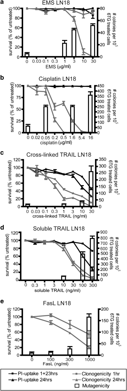
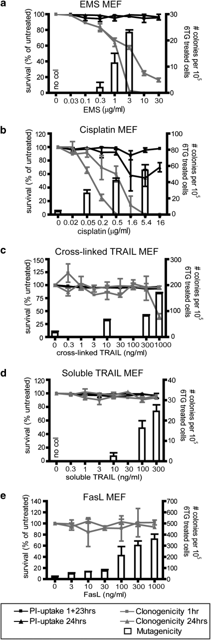
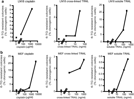
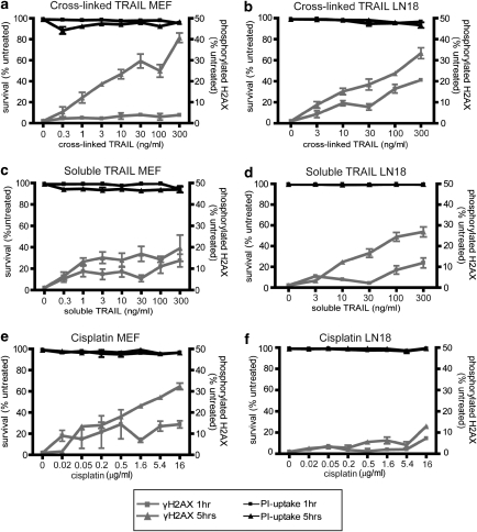
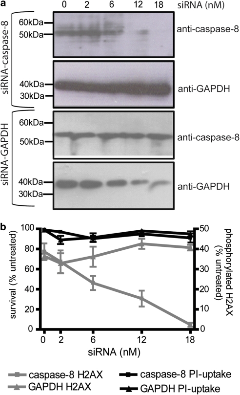
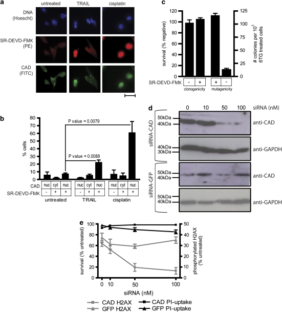
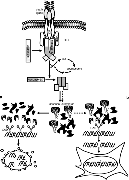
Chemotherapy and radiotherapy commonly damage DNA and trigger p53-dependent apoptosis through intrinsic apoptotic pathways. Two unfortunate consequences of this mechanism are resistance due to blockade of p53 or intrinsic apoptosis pathways, and mutagenesis of non-malignant surviving cells which can impair cellular function or provoke second malignancies. Death ligand-based drugs, such as tumor necrosis factor-related apoptosis inducing ligand (TRAIL), stimulate extrinsic apoptotic signaling, and may overcome resistance to treatments that induce intrinsic apoptosis. As death receptor ligation does not damage DNA as a primary mechanism of pro-apoptotic action, we hypothesized that surviving cells would remain genetically unscathed, suggesting that death ligand-based therapies may avoid some of the adverse effects associated with traditional cancer treatments. Surprisingly, however, treatment with sub-lethal concentrations of TRAIL or FasL was mutagenic. Mutations arose in viable cells that contained active caspases, and overexpression of the caspase-8 inhibitor crmA or silencing of caspase-8 abolished TRAIL-mediated mutagenesis. Downregulation of the apoptotic nuclease caspase-activated DNAse (CAD)/DNA fragmentation factor 40 (DFF40) prevented the DNA damage associated with TRAIL treatment. Although death ligands do not need to damage DNA in order to induce apoptosis, surviving cells nevertheless incur DNA damage after treatment with these agents.


Figures

Figure 1
Figure 1
TRAIL or FasL treatment of LN18 cells provokes generation of 6-TG-resistant colonies. LN18 cells were incubated with various doses of ethane methyl sulfonate (a), cisplatin (b), cross-linked TRAIL (c), soluble TRAIL (d) or Fas ligand (e), either for 24 h or for 1 h followed by 23 h incubation in normal media. Propidium iodide uptake (ad) or clonogenicity assays (ae) were then performed, as outlined in the Materials and methods. Cells were exposed to selected doses of the drugs for 1 h for HPRT mutagenicity assays. The left Y-axis and black and gray lines depict survival as measured in acute cell death and clonogenicity assays. The right Y-axis and white columns show the number of 6-TG-resistant colonies in mutational assays. Error bars indicate standard errors of the means from either three (a, b, e), four (c) or five (d) independent experiments.
Figure 2
Figure 2
TRAIL or FasL treatment of mouse embryo fibroblast (MEF) cells provokes generation of 6-TG-resistant colonies. MEF cells were incubated with various doses of ethane methyl sulphonate (a), cisplatin (b), cross-linked TRAIL (c), soluble TRAIL (d) or Fas ligand (e), for 1hr or 24 h, prior to propidium iodide uptake (ad) or clonogenicity assays (ae). Cells were exposed to selected doses of the drugs for 1 h for HPRT mutagenicity assays. The left Y-axis and black and gray lines depict survival as measured in acute cell death and clonogenicity assays. The right Y-axis and white columns show the number of 6-TG-resistant colonies in mutational assays. Doses, which failed to generate any 6-TG-resistant colonies are labeled ‘no col'. Error bars indicate standard errors of the means from either three (a, b, e), four (c) or five (d) independent experiments.
Figure 3
Figure 3
Mutation frequencies of TRAIL and cisplatin. Mean mutation frequencies of LN18 (a) and mouse embryo fibroblast (b) cells are expressed as the percentage of clonogenically competent cells that generated 6-TG-resistant colonies. The X-axes show the doses analyzed (in log scale). Only doses that enabled the clonogenic survival of more than five cells per experiment were included in this analysis.
Figure 4
Figure 4
TRAIL treatment stimulates phosphorylation of H2AX. The right Y-axes and gray lines show the proportion of MEF cells (a, c, e) or LN18 cells (b, d, f) bearing phosphorylated H2AX after 1 or 5 h exposure to cross-linked TRAIL (a, b), soluble TRAIL (c, d) or cisplatin (e, f). The left Y-axes and black lines represent the percentage of viable cells after same drug exposures. Error bars indicate s.e.m. from three independent experiments.
Figure 5
Figure 5
CrmA expression prevents the formation of 6-TG-resistant mouse embryo fibroblast (MEF) colonies following TRAIL treatment. MEF cells were stably transfected with empty vector (pEF), a wild-type FLAG-tagged crmA expression plasmid or a loss-of-function FLAG-crmA expression plasmid. Genomic DNA from pEF clones was amplified using PCR (a). Products of reactions containing either no template or 100 ng of plasmid are denoted ‘pos cont' and ‘neg cont' respectively. CrmA expression was analyzed using anti-FLAG immunoblotting, relative to a GAPDH loading control (b). (c) The phenotypes of crmA wild-type and mutant clones were tested by transiently transfecting the clones with empty vector (pEF), crmA and/or caspase-8 expression plasmids (90% of transfected DNA), along with a β-galactosidase expression plasmid (10%). The transfectants were stained with Xgal and the blue (transfected) cells were scored visually for morphological characteristics of apoptosis. The proportions of each transiently transfected plasmid are indicated under the graph, as are the stable clones into which this DNA was introduced. Clonogenic survival of each clone was monitored following 1 h exposure to cross-linked TRAIL (d) or cisplatin (f). Parental MEF cells and the indicated stable cell lines were treated with cross-linked TRAIL(e) or cisplatin (g) for 1 h then subjected to the HPRT mutational assay, and the 6-TG-resistant clones that emerged were counted.
Figure 6
Figure 6
CrmA expression prevents TRAIL-induced DNA damage in LN18 cells. (a) LN18 cells were transiently transfected with the indicated plasmids (90% of transfected DNA), along with a β-galactosidase expression plasmid (10%). The transfectants were stained with Xgal and the blue (transfected) cells were scored visually for apoptosis based on morphological criteria. The percentage of the transiently transfected plasmids are indicated under the graph. (b) LN18 cells were transiently transfected with an empty vector (pEF) or plasmids encoding wild-type or mutant crmA, then incubated with the indicated doses of cross-linked TRAIL, soluble TRAIL or cisplatin for 5 h. The proportions of cells bearing phosphorylated H2AX were quantitated by flow cytometry. Error bars indicate standard errors of the means from three independent experiments.
Figure 7
Figure 7
Downregulation of caspase-8 prevents TRAIL-induced DNA damage. MEF cells were transfected with the indicated concentrations of siRNAs targeting caspase-8 or glyceraldehyde 3-phosphate dehydrogenase then subjected to immunoblotting (a) or treated with 300 ng/ml cross-linked TRAIL and assayed for H2AX phosphorylation (b). The left Y-axis and black lines represent the percentage of viable cells. The right Y-axis and gray lines show the proportion of cells bearing phosphorylated H2AX. Error bars indicate s.e.m. from three independent experiments.
Figure 8
Figure 8
Caspase-activated CAD is required for TRAIL to provoke mutations in surviving cells. (a) Fluorescent microscopy was used to investigate CAD localization and SR-DEVD-FMK binding, as a measure of caspase-3/7 activation, in untreated MEF cells or cells incubated for 24 h with cisplatin (5.4 μg/ml) or cross-linked TRAIL (300 ng/ml). The scale bar is 100 μm. (b) The proportions of cells bearing nuclear CAD or SR-DEVD-FMK fluorescence after each treatment were counted. At least 250 cells were scored for each treatment per experiment for untreated and TRAIL-treated samples; at least 100 cisplatin-treated cells were scored per experiment. Error bars indicate s.e.m. from four, three or seven replicates of untreated, cisplatin-treated or TRAIL-treated cells respectively. P-values were calculated using a Student's t-test. (c) After exposure to 300 ng/ml cross-linked TRAIL for 24 h, SR-DEVD-FMK positive and negative cells were flow cytometrically sorted and their clonogenicity assayed following treatment with 300 ng/ml cross-linked TRAIL for 1 h. SR-DEVD-FMK positive and negative sorted cells were also subjected to the HPRT mutational assay, and the 6-TG-resistant clones that emerged were counted. Error bars show the s.e.m. from six or nine independent experiments for clonogenicity and mutagenicity experiments respectively. MEF cells were transfected with the indicated concentrations of small interfering RNAs targeting CAD or GFP then (d) subjected to immunoblotting or (e) treated with 300 ng/ml cross-linked TRAIL and assayed for H2AX phosphorylation. The left Y-axis and black lines represent the percentage of viable cells. The right Y-axis and gray lines show the proportion of cells bearing phosphorylated H2AX. Error bars indicate s.e.m. from three independent experiments.
Figure 9
Figure 9
TRAIL-induced mutagenesis model. Death receptor ligation promotes formation of the death inducing signalling complex (DISC) and activation of caspase-8, which then activates caspases-3 and/or -7. This can trigger apoptosis (a), through cleavage of numerous cellular substrates including ICAD, whose cleavage provokes activation of CAD which fragments DNA. Data reported herein suggest that in cells bearing only a small number of activated effector caspases, limited caspase-mediated ICAD proteolysis leads to CAD activation and DNA cleavage, but not apoptosis (b). We suggest that inaccurate repair of these lesions creates mutations in surviving cells.

Similar articles

Cited by

References

    1. Abraham MC, Shaham S. Death without caspases, caspases without death. Trends Cell Biol. 2004;14:184–193. - PubMed
    1. Ashkenazi A. Targeting the extrinsic apoptosis pathway in cancer. Cytokine Growth Factor Rev. 2008;19:325–331. - PubMed
    1. Ashkenazi A, Pai RC, Fong S, Leung S, Lawrence DA, Marsters SA, et al. Safety and antitumor activity of recombinant soluble Apo2 ligand. J Clin Invest. 1999;104:155–162. - PMC - PubMed
    1. Ashley DM, Riffkin CD, Lovric MM, Mikeska T, Dobrovic A, Maxwell JA, et al. In vitro sensitivity testing of minimally passaged and uncultured gliomas with TRAIL and/or chemotherapy drugs. Br J Cancer. 2008;99:294–304. - PMC - PubMed
    1. Brunelle JK, Letai A. Control of mitochondrial apoptosis by the Bcl-2 family. J Cell Sci. 2009;122:437–441. - PMC - PubMed

Publication types

MeSH terms