Review
. 2010 May 27;11(6):2306-21.
doi: 10.3390/ijms11062306.

An antiapoptotic neuroprotective role for neuroglobin

Affiliations
Review

An antiapoptotic neuroprotective role for neuroglobin

Thomas Brittain et al. Int J Mol Sci. .

Abstract

Cell death associated with mitochondrial dysfunction is common in acute neurological disorders and in neurodegenerative diseases. Neuronal apoptosis is regulated by multiple proteins, including neuroglobin, a small heme protein of ancient origin. Neuroglobin is found in high concentration in some neurons, and its high expression has been shown to promote survival of neurons in vitro and to protect brain from damage by both stroke and Alzheimer's disease in vivo. Early studies suggested this protective role might arise from the protein's capacity to bind oxygen or react with nitric oxide. Recent data, however, suggests that neither of these functions is likely to be of physiological significance. Other studies have shown that neuroglobin reacts very rapidly with cytochrome c released from mitochondria during cell death, thus interfering with the intrinsic pathway of apoptosis. Systems level computational modelling suggests that the physiological role of neuroglobin is to reset the trigger level for the post-mitochondrial execution of apoptosis. An understanding of the mechanism of action of neuroglobin might thus provide a rational basis for the design of new drug targets for inhibiting excessive neuronal cell death.

Keywords: apoptosis; cytochrome c; mitochondria; neuroglobin; systems biology.


Figures
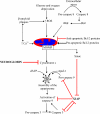
Figure 1
Figure 1
The intrinsic pathway of apoptosis. Mitochondrial pathway of cell death is triggered by a number of internal and external stimuli, and heavily regulated by proteins both up-stream and down-stream of mitochondria. Pre-mitochondrial events that regulate mitochondrial outer membrane permeabilisation (MOMP) in neurons include increase in Ca2+ and reactive oxygen species (ROS) levels. MOMP is tightly regulated by the multi-protein Bcl-2 family, which consists of both anti- and pro-apoptotic members. Down-stream of mitochondria, the pathway is regulated by the low-probability event of apoptosome assembly, as well as positive and negative feedback loops involving caspase 3 and 9, and inhibitor of apoptosis proteins such as XIAP. Cytochrome c is a key protein, initiating apoptosome assembly and activation of caspase cascade. Neuroglobin binding to and reduction of cytochrome c interferes with the mitochondrial pathway of apoptosis immediately down-stream of mitochondria, affecting all types of up-stream stress signals. Lines in red represent inhibitory effects.
Figure 2
Figure 2
Neuroglobin shares little amino acid sequence homology with other family members and is highly conserved throughout species. Protein alignment of neuroglobin and (a) other human globin family members or (b) showing homology between species. Residues marked in red match the consensus, residues in blue are weakly conserved. Protein alignments were performed using MultAlin ver 5.4.1 [15]. (c) Phylogenetic tree representing conservation of neuroglobin throughout evolution. Phylogenetic analysis was performed using Phylogeny.fr. [16].
Figure 2
Figure 2
Neuroglobin shares little amino acid sequence homology with other family members and is highly conserved throughout species. Protein alignment of neuroglobin and (a) other human globin family members or (b) showing homology between species. Residues marked in red match the consensus, residues in blue are weakly conserved. Protein alignments were performed using MultAlin ver 5.4.1 [15]. (c) Phylogenetic tree representing conservation of neuroglobin throughout evolution. Phylogenetic analysis was performed using Phylogeny.fr. [16].
Figure 3
Figure 3
Cytochrome c binding to neuroglobin. (a) Shows the ensemble of complexes calculated from soft docking calculations. The centres of mass of the docked cytochrome c molecules are indicated by the small spheres. (b) Shows the structure of the lowest energy docked complex with neuroglobin in blue and cytochrome c in green.
Figure 4
Figure 4
A co-ordinating role for neuroglobin. The diagram shows the three actions of neuroglobin: (1) in suppressing apoptosis by binding and reducing cytochrome c released from mitochondria, (2) in inhibiting the production of IP3 by GPCR and (3) maintaining the auto-feedback loop, which limits the release of calcium from endoplasmic reticulum (ER) via type 2 IP3 receptors by binding cytochrome c.

References

    1. Galluzzi L, Blomgren K, Kroemer G. Mitochondrial membrane permeabilization in neuronal injury. Nat. Rev. Neurosci. 2009;10:481–494. - PubMed
    1. Lin MT, Beal MF. Mitochondrial dysfunction and oxidative stress in neurodegenerative diseases. Nature. 2006;443:787–795. - PubMed
    1. Skommer J, Wlodkowic D, Deptala A. Larger than life: Mitochondria and the Bcl-2 family. Leuk. Res. 2007;31:277–286. - PubMed
    1. Liston P, Fong WG, Korneluk RG. The inhibitors of apoptosis: There is more to life than Bcl2. Oncogene. 2003;22:8568–8580. - PubMed
    1. Lankester ER. A contribution to the knowledge of haemoglobion. Proc. Roy. Soc. 1872;21:70–81.

Publication types

MeSH terms

LinkOut - more resources