Comparative Study
. 2010 Jul 12;2(3):e00037.
doi: 10.1042/AN20100010.

Dual induction of TREM2 and tolerance-related transcript, Tmem176b, in amyloid transgenic mice: implications for vaccine-based therapies for Alzheimer's disease

Affiliations
Comparative Study

Dual induction of TREM2 and tolerance-related transcript, Tmem176b, in amyloid transgenic mice: implications for vaccine-based therapies for Alzheimer's disease

Benoit Melchior et al. ASN Neuro. .

Abstract

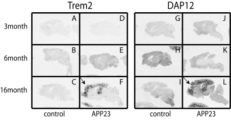
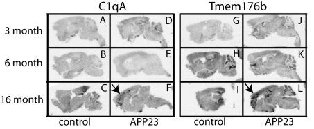
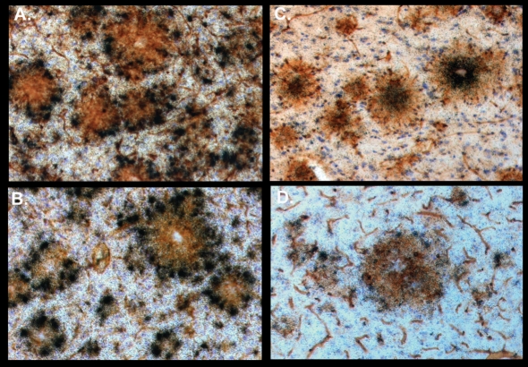
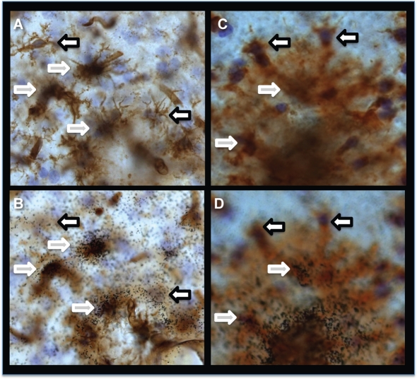
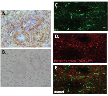
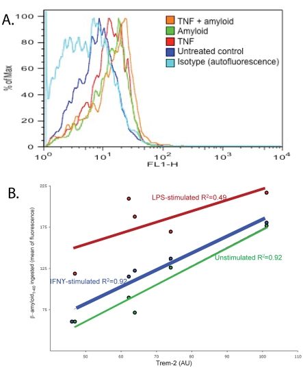
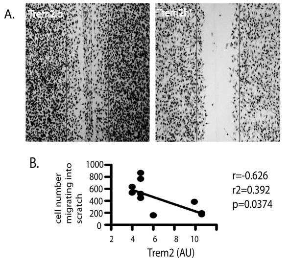
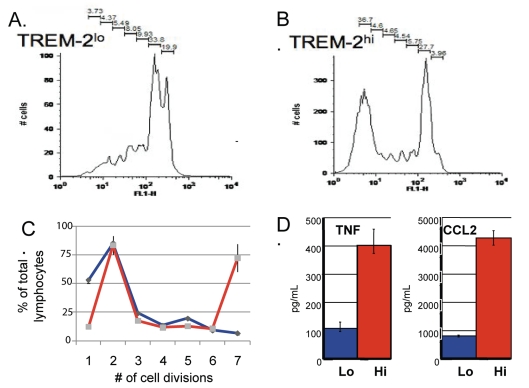
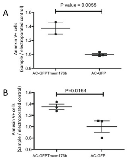
Vaccine-based autoimmune (anti-amyloid) treatments are currently being examined for their therapeutic potential in Alzheimer's disease. In the present study we examined, in a transgenic model of amyloid pathology, the expression of two molecules previously implicated in decreasing the severity of autoimmune responses: TREM2 (triggering receptor expressed on myeloid cells 2) and the intracellular tolerance-associated transcript, Tmem176b (transmembrane domain protein 176b). In situ hybridization analysis revealed that both molecules were highly expressed in plaque-associated microglia, but their expression defined two different zones of plaque-associated activation. Tmem176b expression was highest in the inner zone of amyloid plaques, whereas TREM2 expression was highest in the outer zone. Induced expression of TREM2 occurred co-incident with detection of thioflavine-S-positive amyloid deposits. Transfection studies revealed that expression of TREM2 correlated negatively with motility, but correlated positively with the ability of microglia to stimulate CD4(+) T-cell proliferation, TNF (tumour necrosis factor) and CCL2 (chemokine ligand 2) production, but not IFNgamma (interferon gamma) production. TREM2 expression also showed a positive correlation with amyloid phagocytosis in unactivated cells. However, activating cells with LPS (lipopolysaccharide), but not IFNgamma, reduced the correlation between TREM2 expression and phagocytosis. Transfection of Tmem176b into both microglial and macrophage cell lines increased apoptosis. Taken together, these data suggest that, in vivo, Tmem176b(+) cells in closest apposition to amyloid may be the least able to clear amyloid. Conversely, the phagocytic TREM2(+) microglia on the plaque outer zones are positioned to capture and present self-antigens to CNS (central nervous system)-infiltrating lymphocytes without promoting pro-inflammatory lymphocyte responses. Instead, plaque-associated TREM2(+) microglia have the potential to evoke neuroprotective immune responses that may serve to support CNS function during pro-inflammatory anti-amyloid immune therapies.

Keywords: Aβ, amyloid β peptide; CCL2, chemokine ligand 2; CFSE, carboxyfluorescein succinimidyl ester; CNS, central nervous system; Clast1; DAMP, danger-associated molecular pattern; DMEM, Dulbecco's modified Eagle's medium; EAE, experimentally induced autoimmune encephalomyelitis; FBS, fetal bovine serum; GFP, green fluorescent protein; HPRT, hypoxanthine phosphoribosyl transferase; IFNγ, interferon γ; IL, interleukin; KO, knockout; LPS, lipopolysaccharide; PFA, paraformaldehyde; TNF, tumour necrosis factor; TREM2, triggering receptor expressed on myeloid cells 2; Thio-S, thioflavine-S; Tmem176b, transmembrane domain protein 176b; Torid; WT, wild-type; antigen presentation; autoimmunity; neuroinflammation; qPCR, quantitative PCR.


Figures

Figure 1
Figure 1. TREM2 and DAP12 expression is induced co-ordinate with amyloid plaque deposition
Autoradiograms of sagittal brain sections from non-transgenic (AC and GI) and APP23 transgenic (DF and JL) age-matched siblings. (AF) Brain sections hybridized with 33P-labelled TREM2 riboprobes. (GL) Brain sections hybridized with 33P-labelled DAP12 riboprobes. Arrows indicated amyloid-associated induction of TREM2 and DAP12 expression.
Figure 2
Figure 2. Induced TREM2 expression surrounds Thio-S-positive plaques
(A and B) A brain section from a 10-month-old APP23 transgenic mouse. A compact parenchymal plaque is visualized in green with Thio-S (A). TREM2 expression is visualized with a 33P-labelled ribroprobe (white grains in film emulsion) (B).
Figure 3
Figure 3. C1qA and Tmem176b expression increases in WT mice with age and in association with amyloid plaque deposition in APP23 transgenic mice
Autoradiograms of sagittal brain sections from non-transgenic (AC and GI) and APP23 transgenic (DF and JL) age-matched siblings. (AF) Brain sections hybridized with 33P-labelled C1qA riboprobes. (GL) Brain sections hybridized with 33P-labelled Tmem176b riboprobes. Arrows indicated amyloid-associated induction of C1qA and Tmem176b expression.
Figure 4
Figure 4. Different zones of amyloid-associated microglial activation are defined by regional gene expression
(AD) Brain sections from 16-month-old APP23 transgenic mice. Microglia, macrophages and blood vessels are visualized in brown with tomato lectin. Nuclei are visualized in blue with haematoxylin. Expression of TREM2 (A), DAP12 (B), Tmem176b (C) and C1qA (D) are visualized by 33P-labelled ribroprobes (black grains in film emulsion).
Figure 5
Figure 5. TREM2 and Tmem176b are expressed by plaque-associated microglia in the outer and inner zones respectively of the amyloid plaque
(AD) Brain sections from 16-month-old APP23 transgenic mice. Microglia, macrophages and blood vessels are visualized in brown with tomato lectin. Nuclei are visualized in blue with haematoxylin. Expression of TREM2 (A and B) and Tmem176b (C and D) are visualized by 33P-labelled ribroprobes (black grains in film emulsion). (A and C) The focus at the level of the tomato lectin labelling. (B and D) The focus at the level of the film emulsion. Left-pointing arrows indicate examples of tomato-lectin-positive cells negative for riboprobe labelling. Right-pointing arrows indicate examples of tomato-lectin-positive cells positive for riboprobe labelling.
Figure 6
Figure 6. TREM2 binding activity is detected in the region of amyloid plaque deposition TREM2 expression is induced by aggregated amyloid and promotes amyloid phagocytosis
TREM2–IgG fusion protein labelling of APP23 brain sections is visualized in brown by DAB (diaminobenzidine) immunohistochemistry (A) and by immunofluorescence in red (D and E). Microglia and macrophages are visualized by immunofluorescence with Iba1 in green (C and E). In (A and B) nuclei are visualized in blue with haematoxylin. (B) The absence of immunolabelling with secondary antibodies directed against human IgG. (E) The merged images of (C and D).
Figure 7
Figure 7. TREM2 expression is induced by aggregated amyloid and promotes amyloid phagocytosis
(A) Flow-cytometric analysis of TREM2 expression of the surface of CD11b+ microglia in primary mixed glial cultures treated for 5 days with TNF only, amyloid only or both TNF and amyloid. (B) Flow-cytometric analysis of cloned BV-2 microglial cell lines phagocytosis of fluorescently labelled AB1-40 (mean fluorescence intensity relative to WT BV-2 cells compared with TREM2 surface expression) in untreated (green line), IFNγ-treated (blue line) and LPS-treated (red) cell lines. APC (allophycocyanin)-labelled TREM2 antibodies were used to quantify the level of TREM2 expression in amyloid-treated BV-2 cell lines.
Figure 8
Figure 8. TREM2 expression negatively correlates with cell motility
The number of cells within the scratch zone was quantified as a function of TREM2 expression per culture 6 h post-injury. (A) Representative migration in scratched confluent TREM2lo (left-hand side) and TREM2hi (right-hand side) cultures. (B) Number of cells per scratch zone as a function of TREM2 mRNA of each scratch-tested culture. The Pearson correlation co-efficient was used to determine significance.
Figure 9
Figure 9. TREM2 expression positively correlates with antigen presentation
Cloned BV-2 microglial cell lines with ∼10-fold difference in TREM2 RNA expression were used to stimulate the proliferation of allogeneic (BALB/c) CFSE-labelled CD4+ T-cells. Representative flow-cytometric analysis of T-cell proliferation using TREM2lo BV-2 cells or when using TREM2hi are depicted in (A) and (B) respectively. (C) The mean number of cells±S.E.M. undergoing each round of cell division in T-cells stimulated with TREM2lo (blue line) or TREM2hi (red line) BV-2 cells. (D) The levels of TNF and CCL2 found in the supernatants of T-cell cultures stimulated with TREM2 lo (blue bars) or TREM2hi (red bars).
Figure 10
Figure 10. Tmem176b overexpression promotes annexin V labelling
RAW macrophage (A) and BV-2 microglial (B) cell lines were transiently transfected with Tmem176b–GFP overexpression or GFP-only overexpression plasmids. Annexin V labelling of GFP+ cells was determined 48 h post-transfection using flow-cytometric analysis (means±S.E.M. for three replicate experiments). Between replicate experiments, the final total cell death ranged from ∼35 to ∼50% of cultured cells. Significance was determined using the Student's t test.

References

    1. Afagh A, Cummings BJ, Cribbs DH, Cotman CW, Tenner AJ. Localization and cell association of C1q in Alzheimer's disease brain. Exp Neurol. 1996;138:22–32. - PubMed
    1. Bianchin MT, Capella HM, Chaves DL, Steindel M, Grisard EC, Ganev GG, da Silva Júnior JP, Neto Evaldo S, Poffo MA, Walz R, Carlotti Júnior CG, Sakamoto AC. Nasu-Hakola disease (polycystic lipomembranous osteodysplasia with sclerosing leukoencephalopathy–PLOSL): a dementia associated with bone cystic lesions. From clinical to genetic and molecular aspects. Cell Mol Neurobiol. 2004;24:1–24. - PMC - PubMed
    1. Bouchon A, Hernández-Munain C, Cella M, Colonna M. A DAP12-mediated pathway regulates expression of CC chemokine receptor 7 and maturation of human dendritic cells. J Exp Med. 2001;194:1111–1122. - PMC - PubMed
    1. Brynskikh A, Warren T, Zhu J, Kipnis J. Adaptive immunity affects learning behavior in mice. Brain Behav Immun. 2008;22:861–869. - PubMed
    1. Butovsky O, Bukshpan S, Kunis G, Jung S, Schwartz M. Microglia can be induced by IFN-γ or IL-4 to express neural or dendritic-like markers. Mol Cell Neurosci. 2007;35:490–500. - PubMed

Publication types

MeSH terms