Review
. 2010 Apr;2(4):769-90.
doi: 10.4155/bio.10.31.

Immunoaffinity chromatography: an introduction to applications and recent developments

Affiliations
Review

Immunoaffinity chromatography: an introduction to applications and recent developments

Annette C Moser et al. Bioanalysis. 2010 Apr.

Abstract

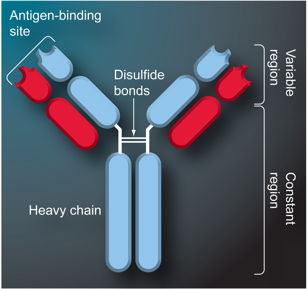
Immunoaffinity chromatography (IAC) combines the use of LC with the specific binding of antibodies or related agents. The resulting method can be used in assays for a particular target or for purification and concentration of analytes prior to further examination by another technique. This review discusses the history and principles of IAC and the various formats that can be used with this method. An overview is given of the general properties of antibodies and of antibody-production methods. The supports and immobilization methods used with antibodies in IAC and the selection of application and elution conditions for IAC are also discussed. Several applications of IAC are considered, including its use in purification, immunodepletion, direct sample analysis, chromatographic immunoassays and combined analysis methods. Recent developments include the use of IAC with CE or MS, ultrafast immunoextraction methods and the use of immunoaffinity columns in microanalytical systems.


Figures

Figure 1
Figure 1. Typical structure of an IgG-class antibody
IgG-class antibodies consist of four polypeptide chains that are linked by disulfide bonds to create a ‘Y’-shaped structure with two identical binding sites.
Figure 2
Figure 2. Process for polyclonal antibody production
Figure adapted with permission from [1]. © Taylor and Francis CRC Press (2006).
Figure 3
Figure 3. Process for monoclonal antibody production
Figure adapted from [1]. © Taylor and Francis CRC Press (2006).
Figure 4
Figure 4. General procedure for immobilizing antibodies within a monolithic column
Reproduced with permission from [18]. © American Chemical Society (2005).
Figure 5
Figure 5. Typical format in which a sample containing the analyte is applied to an immunoaffinity column and nonretained sample components are allowed to pass through
The analyte is later eluted by disrupting the antibody–antigen interactions with an appropriate elution buffer. This on/off mode can be used for direct detection and/or purification of the analyte.
Figure 6
Figure 6. System for combining immunoaffinity chromatography with reversed-phase chromatography
An antibody column is used to extract or preconcentrate analytes from a sample prior to separation of these analytes using a reversed-phase column. This particular system has been used to measure virginiamycin in water samples with a LOD of 1 ppb. RP: Reversed-phase. Figure reproduced with permission from [Carlson T, Moser AC, Unpublished Data].
Figure 7
Figure 7. Simultaneous injection immunoassay format
In this type of competitive immunoassay, the sample and a labeled analog of the analyte are mixed and injected onto an immunoaffinity column. The labeled analog and analyte can bind to this column while other sample components pass through nonretained. The analyte and labeled analog are then eluted with an appropriate mobile phase/elution buffer. In this method the analyte concentration in the sample is inversely related to the amount of retained labeled analog that is detected.
Figure 8
Figure 8. Sequential injection immunoassay format
In this format, the sample is applied onto an immunoaffinity column and the analyte is allowed to bind. A labeled analog of the analyte is then injected onto the same column and also allowed to bind to any remaining free antibody sites. An elution buffer is used to later remove both the retained analyte and labeled analog for the column. The amount of retained labeled analog will be inversely related to the amount of analyte that was in the original sample.
Figure 9
Figure 9. Displacement immunoassay format
In this type of competitive binding immunoassay, a labeled analog of the analyte is injected onto an immunoaffinity column. A sample is then injected onto the column and the analyte is allowed to displace some of the labeled analog. The size of the peak for the displaced label is directly related to the amount of analyte that was in the sample.
Figure 10
Figure 10. Sandwich immunoassay format
In this type of noncompetitive binding immunoassay, the sample is injected onto an immunoaffinity column and the analyte allowed to bind to the immobilized antibodies. A labeled antibody that is specific for the analyte is then injected onto the same column and also allowed to bind, creating a sandwich immune complex for the analyte. An elution buffer is applied to disrupt the antigen–antibody binding and regenerate the column. The amount of retained, labeled antibody that is eluted during this step is directly proportional to the amount of analyte that was present in the original sample.
Figure 11
Figure 11. One-site immunometric assay
In this noncompetitive immunoassay format, the analyte and labeled antibodies are mixed and incubated prior to injection onto an immobilized analyte column. The analyte column binds any excess antibodies and the analyte-bound antibodies are eluted in the nonretained fraction, providing a signal that is directly related to the analyte’s original concentration. The column is later regenerated by eluting off the excess labeled antibodies.
Figure 12
Figure 12. General scheme for postcolumn immunodetection
In this detection method, eluent from an HPLC column is directed into a reaction coil along with an excess of labeled antibodies that are able to bind the desired analyte. The excess antibodies bind to an immobilized analog column and the labeled analyte–antibody complexes are monitored as they elute from the analog column, providing a signal that is directly related to the original amount of eluting analyte. Reproduced with permission from [15]. © American Chemical Society (2001).

References

    1. Hage DS, Phillips TM. Chapter 6 Immunoaffinity chromatography. In: Hage DS, editor. Handbook of Affinity Chromatography. NY, USA: Taylor & Francis; 2006. . ▪ Provides an indepth look at immunoaffinity chromatography, with an emphasis on method development, immunoextraction and sample analysis by immunoaffinity chromatography.

    1. Hage DS. Survey of recent advances in analytical applications of immunoaffinity chromatography. J. Chromatogr. B. 1998;715(1):3–28.. ▪ Provides a good overall review of immunoaffinity chromatography and its analytical applications.

    1. Calton GJ. Immunosorbent separations. Meth. Enzymol. 1985;104:381–387. - PubMed
    1. Phillips TM. High performance immunoaffinity chromatography. An introduction. LC Mag. 1985;3:962–972.
    1. D’allesandro G, Sofia F. The adsorption of antibodies from the sera of syphilitics and turburculosis patients. Z. Immunitats. 1935;84:237–250.

Publication types

Substances