. 2010;12(4):R147.
doi: 10.1186/ar3089. Epub 2010 Jul 19.

Liposomal encapsulation enhances and prolongs the anti-inflammatory effects of water-soluble dexamethasone phosphate in experimental adjuvant arthritis

Affiliations

Liposomal encapsulation enhances and prolongs the anti-inflammatory effects of water-soluble dexamethasone phosphate in experimental adjuvant arthritis

Rebecca Anderson et al. Arthritis Res Ther. 2010.

Abstract

Introduction: The objective of this study was to evaluate the efficacy of intravenous (i.v.) injection of liposomally encapsulated dexamethasone phosphate (DxM-P) in comparison to free DxM-P in rats with established adjuvant arthritis (AA). This study focused on polyethylene glycol (PEG)-free liposomes, to minimize known allergic reactions caused by neutral PEG-modified (PEG-ylated) liposomes.

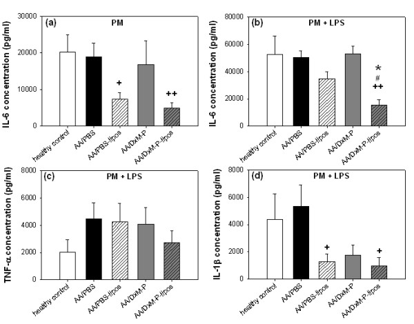
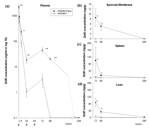
Methods: Efficacy was assessed clinically and histologically using standard scores. Non-specific and specific immune parameters were monitored. Activation of peritoneal macrophages was analyzed via cytokine profiling. Pharmacokinetics/biodistribution of DxM in plasma, synovial membrane, spleen and liver were assessed via mass spectrometry.

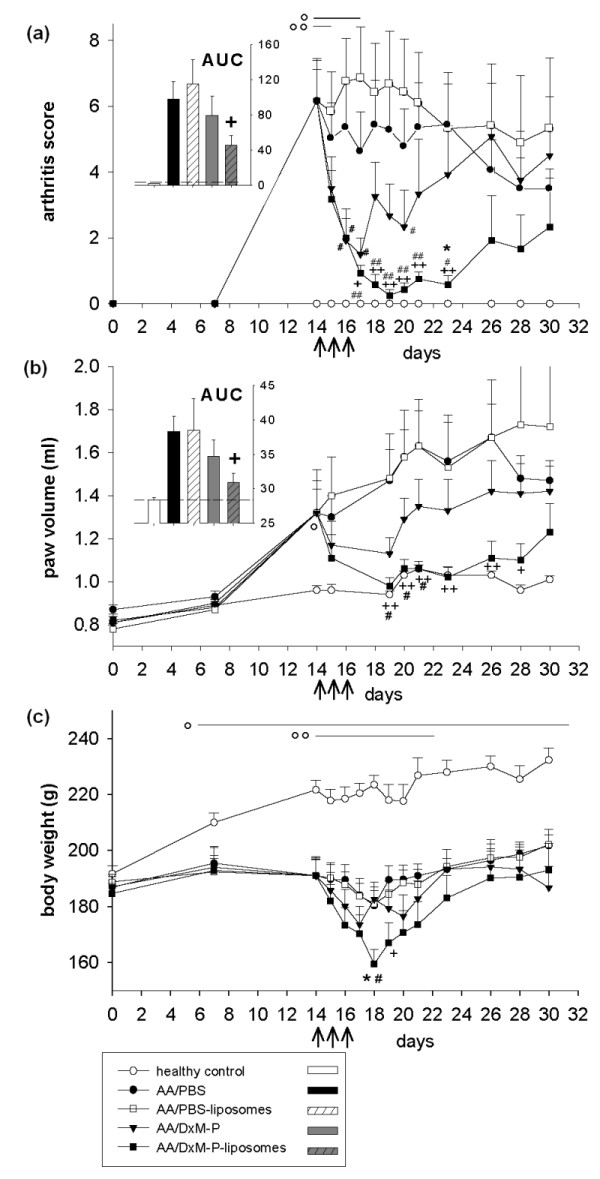
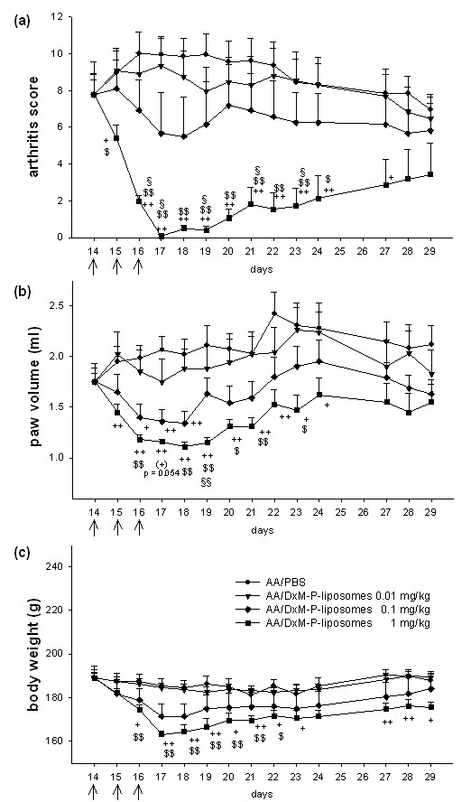
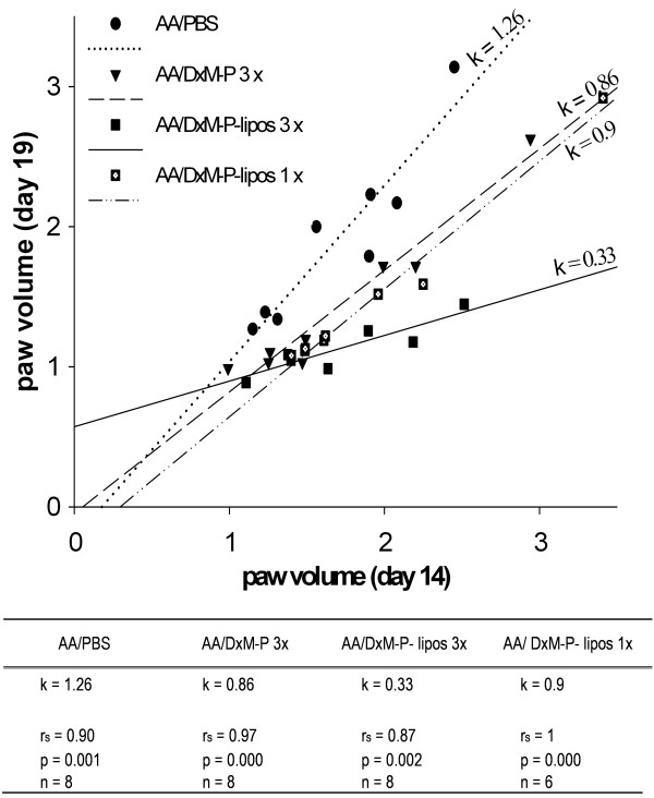
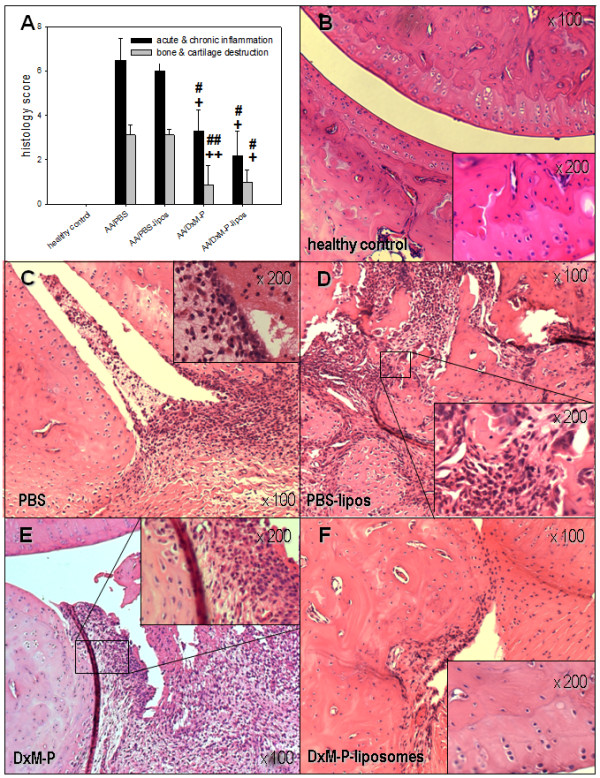
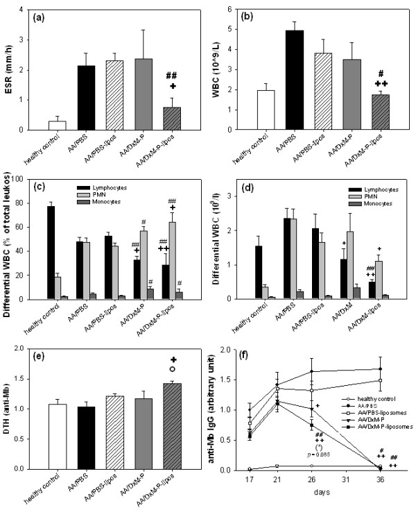
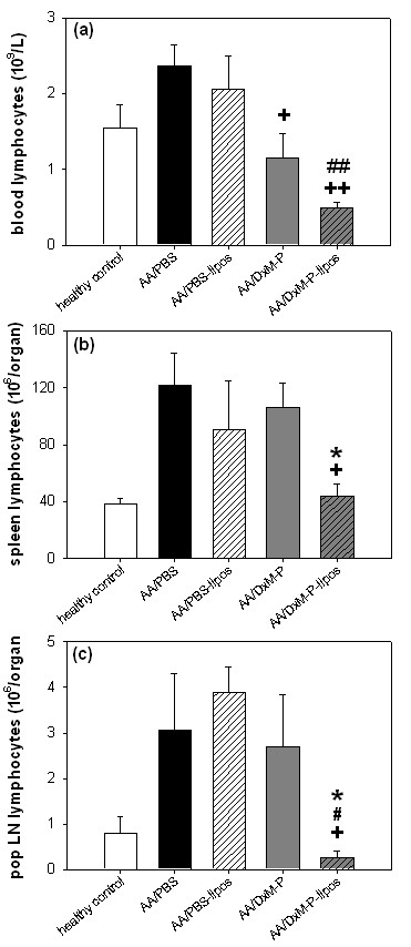
Results: Liposomal DxM-P (3 × 1 mg/kg body weight; administered intravenously (i.v.) on Days 14, 15 and 16 of AA) suppressed established AA, including histological signs, erythrocyte sedimentation rate, white blood cell count, circulating anti-mycobacterial IgG, and production of interleukin-1beta (IL-1β) and IL-6 by peritoneal macrophages. The suppression was strong and long-lasting. The clinical effects of liposomal DxM-P were dose-dependent for dosages between 0.01 and 1.0 mg/kg. Single administration of 1 mg/kg liposomal DxM-P and 3 × 1 mg/kg of free DxM-P showed comparable effects consisting of a partial and transient suppression. Moreover, the effects of medium-dose liposomal DxM-P (3 × 0.1 mg/kg) were equal (in the short term) or superior (in the long term) to those of high-dose free DxM-P (3 × 1 mg/kg), suggesting a potential dose reduction by a factor between 3 and 10 by liposomal encapsulation. For at least 48 hours after the last injection, the liposomal drug achieved significantly higher levels in plasma, synovial membrane, spleen and liver than the free drug.

Conclusions: This new PEG-free formulation of macrophage-targeting liposomal DxM-P considerably reduces the dose and/or frequency required to treat AA, with a potential to enhance or prolong therapeutic efficacy and limit side-effects also in the therapy of rheumatoid arthritis. Depot and/or recirculation effects in plasma, inflamed joint, liver, and spleen may contribute to this superiority of liposomally encapsulated DxM-P.


Figures

Figure 1
Figure 1
Clinical effects of free or liposomal DxM-P in adjuvant arthritis (Experiment 1). Effect of treatment with PBS-liposomes, free DxM-P (3 × 1 mg/kg; Days 14, 15 and 16), or liposomal DxM-P (3 × 1 mg/kg; Days 14, 15 and 16) on arthritis score (a), paw volume (b), and body weight (c); all arthritic groups were normalized to the same mean value for each parameter on Day 14 (n = 6 each). The inserts show the area under the curve (AUC) from Day 0 to Day 30 for the arthritis score (a) and the paw volume (b); dashed line: healthy control levels. For all treatment groups: + P ≤ 0.05, ++ P ≤ 0.01 vs. AA/PBS; # P ≤ 0.05, ## P ≤ 0.01 vs. AA/PBS-liposomes; * P ≤ 0.05 vs. AA/DxM-P; For AA/liposomal DxM-P: o P ≤ 0.05, oo P ≤ 0.01 vs. healthy controls; all Mann Whitney U-test; ↑ I. v. administration of PBS, PBS-liposomes, free DxM-P, or liposomal DxM-P.
Figure 2
Figure 2
Dose-dependence of liposomal DxM-P in adjuvant arthritis (Experiment 4). (a-c): Dose-response of treatment with liposomal DxM-P (3 × 1 mg/kg, 3 × 0.1 mg/kg, or 3 × 0.01 mg/kg); all groups were normalized to the same mean value for each parameter on Day 14 (n = 6 each). + P ≤ 0.05, ++ P ≤ 0.01 vs. AA/PBS; $ P ≤ 0.05, $$ P ≤ 0.01 vs. AA/DxM-P-liposomes 0.01 mg/kg; § P ≤ 0.05, §§ P ≤ 0.01 vs. AA/DxM-P-liposomes 0.1 mg/kg, * P ≤ 0.05 vs. AA/DxM-P 1 mg/kg; all Mann Whitney U-test. ↑ administration of PBS or liposomal DxM-P.
Figure 3
Figure 3
Correlation between the paw volume on Days 14 and 19 during the time course of adjuvant arthritis. Correlation (Spearman rank test, rs) between the paw volume on Day 19 (maximal efficacy of treatment) and Day 14 (prior to treatment) of individual rats treated with PBS (3 ×; Days 14, 15 and 16; n = 8), free DxM-P (3 × 1 mg/kg; Days 14, 15 and 16; n = 8), or liposomal DxM-P (3 × 1 mg/kg; Days 14, 15 and 16; n = 8; or 1 × 1 mg/kg; Day 14; n = 6). The solid, dashed, and dotted lines are the regression lines, k represents the regression gradient.
Figure 4
Figure 4
Histological effects of liposomal DxM-P in adjuvant arthritis (Day 21; Experiment 2/3). Inflammation and destruction score (a) and images of HE-stained joints sections from healthy controls (b) or arthritic rats after treatment with PBS (c), PBS-liposomes (d), free DxM-P (e), or liposomal DxM-P (f) (for details see Figure 1). + P ≤ 0.05, ++ P ≤ 0.01 vs. AA/PBS; # P ≤ 0.05, ## P ≤ 0.01 vs. AA/PBS-liposomes; all Mann Whitney U-test.
Figure 5
Figure 5
Hematological parameters (Day 21) and anti-Mb IgG response (Days 17 to 36) in adjuvant arthritis (Experiment 2/3). Erythrocyte sedimentation rate (ESR) (a), white blood cell count (WBC) (b), differential WBC in relative percentages (c) and absolute numbers of leukocytes (d), delayed type hypersensitivity (DTH) (e), and time course of serum anti-Mb IgG (f) after treatment as in Figure 1; n = 8 for all groups. + P ≤ 0.05, ++ P ≤ 0.01 vs. AA/PBS; # P ≤ 0.05, ## P ≤ 0.01 vs. AA/PBS-liposomes; (*) P ≤ 0.085 vs. AA/DxM-P; all Mann Whitney U-test.
Figure 6
Figure 6
Effects of liposomal DxM-P on lymphocytes in adjuvant arthritis (Day 21; Experiment 2/3). Assessment of lymphocytes in blood (a) (n = 8), spleen (b) (n = 4) and pop LN (c) (n = 4) after treatment with PBS, PBS-liposomes, free DxM-P, or liposomal DxM-P (for details see Figure 1). + P ≤ 0.05, ++ P ≤ 0.01 vs. AA/PBS; # P ≤ 0.05, ## P ≤ 0.01 vs. PBS-liposomes; * P ≤ 0.05 vs. AA/DxM-P; all Mann Whitney U-test.
Figure 7
Figure 7
Cytokine production by peritoneal Mφ in adjuvant arthritis (Day 21; Experiment 2/3). IL-6 (a-b), TNF-α (c), and IL-1β (d) production by non-stimulated and LPS-stimulated peritoneal Mφ after treatment as in Figure 1; n = 4 for all groups. + P ≤ 0.05, ++ P ≤ 0.01 vs. AA/PBS; # P ≤ 0.05 vs. AA/PBS-liposomes; * P ≤ 0.05 vs. AA/DxM-P; all Mann Whitney U-test.
Figure 8
Figure 8
Biodistribution of free or liposomal DxM-P in adjuvant arthritis. Concentration of DxM in blood (a) (n = 6), synovial membrane (b) (n = 3), spleen (c) (n = 3), and liver (d) (n = 3) at 0.5, 2, 8, 24, and/or 72, 96, and 288 hours after the initial i. v. treatment with free or liposomal DxM-P during the time course of AA (for details see Figure 1). * P ≤ 0.05, ** P ≤ 0.01 vs. AA/DxM-P; all Mann Whitney U-test; administration of free or liposomal DxM-P at the time points 0 hours, 24 hours, and 48 hours.

References

    1. Firestein GS, Alvaro-Gracia JM, Maki R. Quantitative analysis of cytokine gene expression in rheumatoid arthritis. J Immunol. 1990;144:3347–3353. - PubMed
    1. Mulherin D, Fitzgerald O, Bresnihan B. Synovial tissue macrophage populations and articular damage in rheumatoid arthritis. Arthritis Rheum. 1996;39:115–124. doi: 10.1002/art.1780390116. - DOI - PubMed
    1. Kinne RW, Stuhlmüller B, Burmester GR. Cells of the synovium in rheumatoid arthritis. Macrophages. Arthritis Res Ther. 2007;9:224. doi: 10.1186/ar2333. - DOI - PMC - PubMed
    1. Schulze-Koops H, Davis LS, Kavanaugh AF, Lipsky PE. Elevated cytokine messenger RNA levels in the peripheral blood of patients with rheumatoid arthritis suggest different degrees of myeloid cell activation. Arthritis Rheum. 1997;40:639–647. doi: 10.1002/art.1780400408. - DOI - PubMed
    1. Bresnihan B. Pathogenesis of joint damage in rheumatoid arthritis. J Rheumatol. 1999;26:717–719. - PubMed

Publication types

MeSH terms